R.Du Hyun RoOrthopedics & Research
Text

RESEARCH TO PRACTICE · CONNECTEVE

Orthopedic research
becomes a tool for care.

Osteoarthritis progresses differently in each person. I study ways to assess the current condition using consistent criteria, record changes quantitatively, and predict what happens next.

Making measurement methods from research usable in the clinic, and explaining their results so patients can understand them, are part of the same work. At CONNECTEVE, we apply this research to the development of medical AI, software, and surgical robots.

Prognosis research

Reading the future course from the knee we see today.

Beyond assessing osteoarthritis, we have studied which knees change faster and which information explains the differences. These four studies examine alignment change, OA progression, onset in the other knee and structural differences within the same grade.

Knee Surgery, Sports Traumatology, Arthroscopy

01 · Knee Surgery, Sports Traumatology, Arthroscopy

How quickly does leg alignment change?

We followed standing long-leg radiographs of 10,841 limbs to examine how baseline alignment and the joint line convergence angle (JLCA) relate to subsequent changes.

Annual progression of at least 0.50° in the same direction occurred in 34.0% of the baseline varus group and 25.7% of the valgus group. Greater JLCA, baseline alignment and higher OA grades were associated with faster change.

The findings support following the whole-leg axis and joint line alongside joint space.
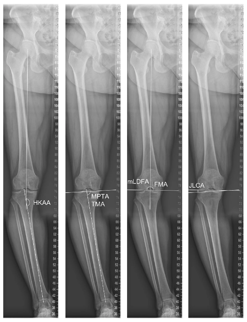
Original Figure 2 · Alignment measurements on standing long-leg radiographs.
DOI · 10.1002/ksa.70293
Original figures and study explanation →
Journal of Orthopaedic Research

02 · Journal of Orthopaedic Research

Can we identify knees likely to progress?

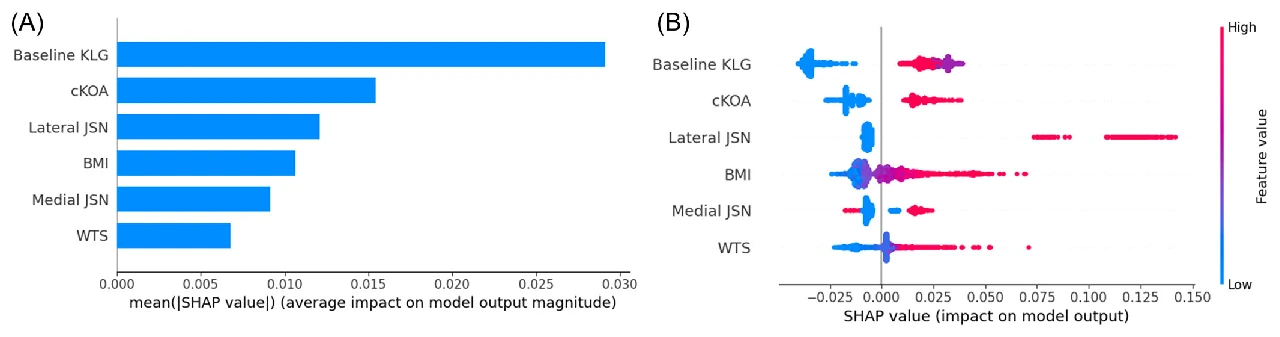
We developed a model using 5,966 OAI knees and tested radiographic progression over 4–5 years in an independent MOST cohort of 3,392 knees.

Six variables were selected: baseline KL grade, contralateral OA, medial and lateral joint space narrowing, BMI and the WOMAC score. External validation AUC was 0.76.

This study stratifies follow-up risk using routinely available information. It did not directly predict pain or the need for surgery.

Original Figure 3 · Predictors of progression and SHAP analysis.
DOI · 10.1002/jor.25982
Original figures and study explanation →
Journal of Orthopaedic Research

03 · Journal of Orthopaedic Research

Will osteoarthritis develop in the other knee?

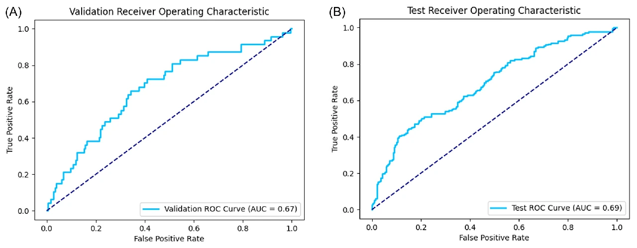
Among 1,353 people with unilateral OA, we used OAI for model development and MOST for independent testing of contralateral OA onset over 4–5 years.

The model used nine variables, including baseline lateral joint space narrowing in the other knee. Independent test AUC was 0.69, indicating moderate discrimination.

The study supports assessing and following the other knee as well as the symptomatic side.

Original Figure 2 · ROC curves in the validation and independent test cohorts.
DOI · 10.1002/jor.26018
Original figures and study explanation →
Osteoarthritis and Cartilage Open

04 · Osteoarthritis and Cartilage Open

Why can knees with the same OA grade follow different courses?

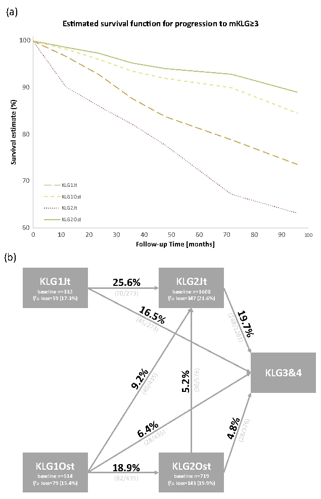
We compared symptoms and progression in 10,804 knees from 5,802 people, separating early KL 1–2 knees into osteophyte-dominant and joint-space-narrowing-dominant phenotypes.

Pain and function did not differ within the same grade, but progression to KL 3 or higher differed by structural phenotype. Osteophyte-dominant knees had a longer mean time to progression.

Structural differences add information beyond a grade number. Radiographic progression and current symptoms require separate assessment.

Original Figure 4 · Progression by early structural phenotype and mean time to progression.
DOI · 10.1016/j.ocarto.2025.100566
Original figures and study explanation →
01 · Objective Assessment

What is the condition of the knee today?

We measure joint space, osteophytes and leg alignment on X-rays in a consistent way. Aligning the measurement items and methods is important so that assessors use the same criteria.

Related papers
02 · Quantification

How much has changed since the last visit?

Add to the arthritis rating and leave spacing and angles as continuous values. We're going to separate the deviation from the actual change in measurement, and we're looking at small changes in tracking observations.

Related papers
03 · Predicting Prognosis

Whose disease may progress faster?

We analyze video and symptoms, life and medical information together to predict future changes. We're also testing performance in other groups and validating the scope of individual treatment decisions.

Related papers

01 · Tools for objective assessment

Review the image findings
together.

To answer “What stage is my arthritis?”, we should explain which features support that assessment. The starting point is consistent assessment of where joint space narrows, where osteophytes lie and the angles of the whole leg.

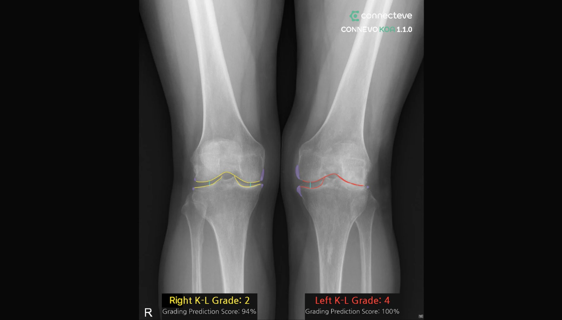
CONNEVO KOA analyzes osteophytes and joint space on knee X-rays and assists K-L grading. Results are displayed on the image for clinicians to review beside the original and use in consultations. AI measures must be interpreted alongside symptoms, examination and imaging conditions.

Example of CONNEVO KOA identifying osteophytes and joint spaces on a knee X-ray
CONNEVO KOA · Osteophyte and joint-space analysis. Grading Prediction Score is an analysis score for the current radiographic grade.

02 · Tools for quantifying structure and change

Record how much
has changed.

Joint space and leg alignment differ even within the same OA grade. Recording change requires repeatable measurements and consideration of positioning differences. My research measures spaces and angles precisely to understand what changes over time.

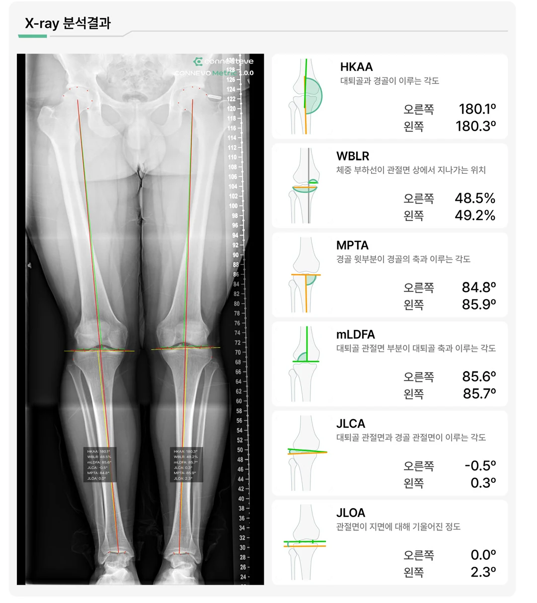
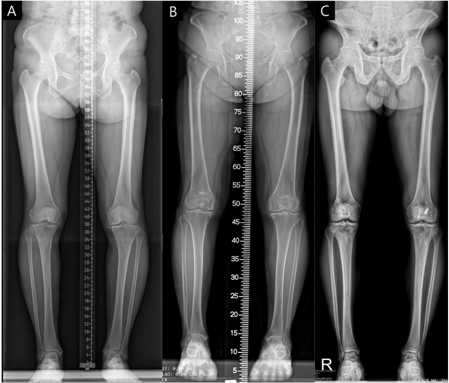
CONNEVO Metric automatically measures alignment on full-length X-rays from hip to ankle. It visualizes HKAA, WBLR, MPTA, mLDFA, JLCA and JLOA and provides a report. These measures create shared information for describing knee structure and reviewing surgical plans.

The work on joint space in OJSW research provides another way to quantify subtle imaging changes. The study evaluated measurement of the minimum joint space perpendicular to the articular surface. oJSW image analysis is used to assess structural change in clinical trials of new drugs for knee osteoarthritis.

X-ray and lower-limb alignment index part of the CONNEVO Metric results
CONNEVO Metric · Lower-limb alignment measurements.

Spine and pelvic alignment · CONNEVO Spin

Understand spine and pelvic alignment

AI measurement of alignment parameters from lateral lumbar-spine X-rays.

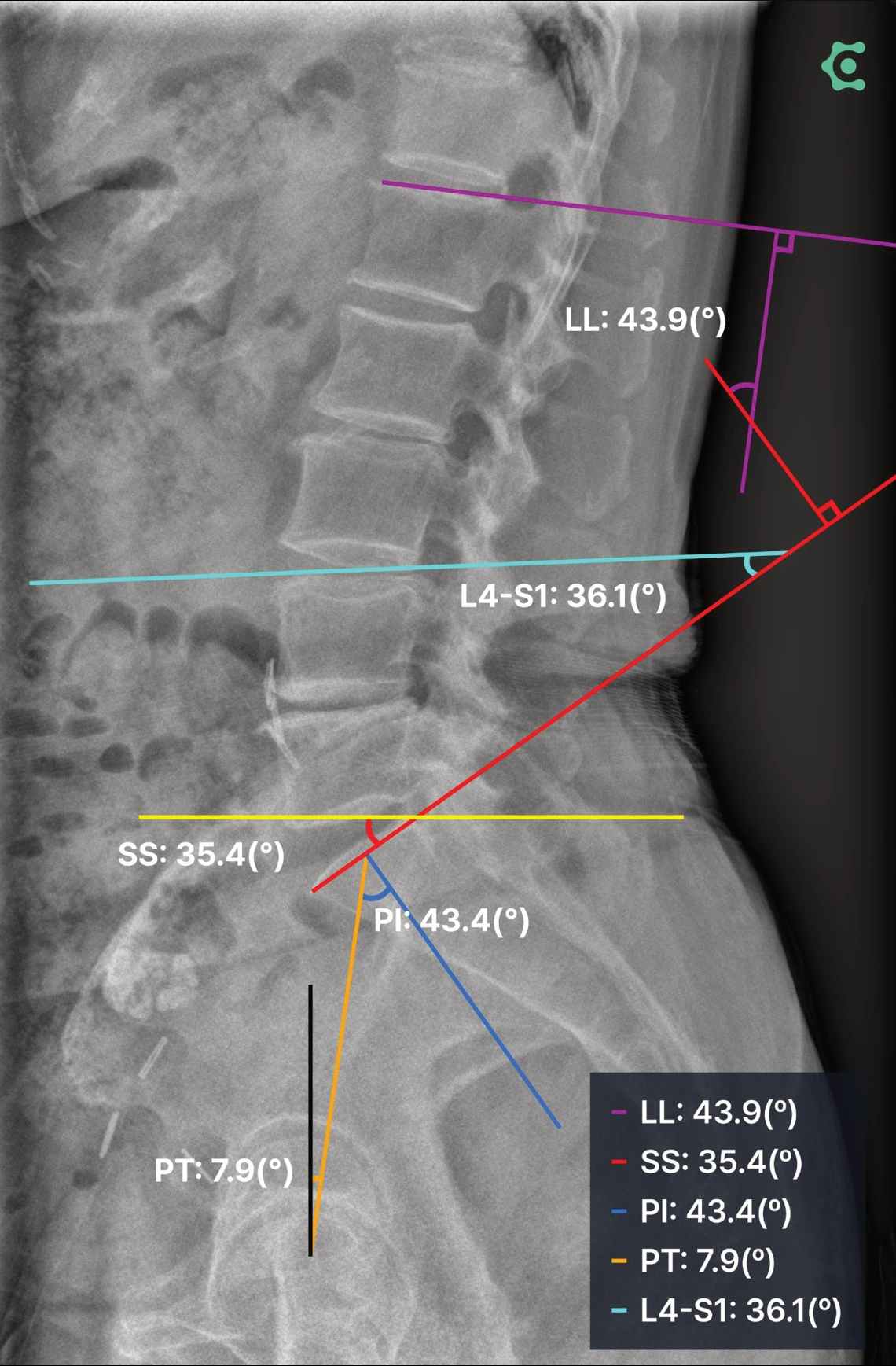
CONNEVO Spin analyzes standing lateral lumbar-spine images to automate alignment measurements, including LL, SS, PI, PT and L4-S1, and supports pre- and postoperative comparison and patient explanation.

Angles and measurements displayed on the X-ray make it easier to follow your clinician’s explanation. Comparing images before and after surgery can help you understand changes in spine and pelvic alignment.

LL
Lumbar lordosis
SS
Sacral slope
PI
Pelvic incidence
PT
Pelvic tilt
L4–S1
Lower lumbar lordosis
CONNEVO Spin measurement example: a lateral lumbar-spine X-ray annotated with LL, SS, PI, PT and L4-S1 angles.
CONNEVO Spin: a spine and pelvic alignment measurement example. The angles shown are measurements from this image.

04 · Explaining the same picture as the patient

Help measured information
helps patients understand their condition.

Understanding what the numbers mean together is as important as obtaining them. Clinicians need to explain what has changed in the images and what each treatment can offer, so patients can ask questions that reflect their lives and goals.

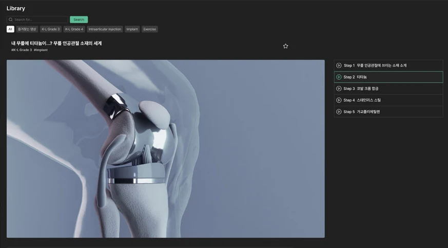
CONNEVO Suite loads KOA and Metric analysis results into the consultation interface and connects them with educational videos, explanatory materials, and patient-specific reports. It supports the explanation of image findings and measurements within the clinical workflow. My patient education content and the exercise and treatment articles on this website share the same purpose.

The CONNEVO Suite has a consultation screen that calls for joint replacement and arthritis educational videos.
CONNEVO Suite · Patient education and consultation.

Surgical planning · Robotics · PHYSICAL AI

Connect imaging information
with movement during surgery.

The study of understanding the structure and planning for knee led to the creation of the surgery robot. We need to figure out what criteria we can use to cut the bone and balance it, how exactly the actual location of surgery matches the plan.

CONNECTEVE is combining CT-based planning with 3D scanning to develop the technology for controlling robots. Physical AI is a study that uses imaging and sensors to understand the real world, how to move your plan into safe movement. Work on building the surgery data and digital twins is also in progress.

Imaging and measurement

Joint space · Alignment · Anatomy

Planning

Individual targets · Bone resection · Balance

The real movement.

3D registration · Sensors · Robot control

Du Hyun Ro’s exercise videos

Watch the movement,
start at your own pace.

Explore strength and stretching exercises for people with arthritis. Adjust the range of motion and exercise volume to your knee condition.

Watch the exercise collection on YouTube

CLINICAL EVIDENCE

From measurement technology to clinical evidence

What was measured and where was it tested? Technology development, external validation and economic modelling answer different questions.

KOA · KNEE OA GRADING

Grading osteoarthritis in a multicentre cohort

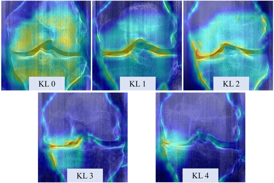
Eigen-CAM · KL 0–4
Lee DW et al. KSRR 2024. Figure 6. Terms

The study evaluated 2,546 knees in 1,273 patients at two Korean university hospitals. For OA detection (KL ≥2), AUC was 0.94 and accuracy 94.2%. KL 2 sensitivity was 77.2%, illustrating the difference between binary OA detection and grade-specific classification. This was standalone AI external validation.

oJSW measures minimum joint space perpendicular to the joint surface. Across 15,313 images, AUCs for JSN >0, >1 and >2 were 0.86, 0.95 and 0.97. These measure discrimination of current radiographic narrowing.

  1. Byung Sun Choi, Seong Hwan Hong, Hae Jung Lee, Seong Hwan Kim. Performance of an Artificial Intelligence-Based Software for Automated Kellgren-Lawrence Grading of Knee Osteoarthritis: A Multicenter Cohort Study. Journal of Arthroplasty · 2026. doi:10.1016/j.arth.2026.01.078.

  2. Do Weon Lee, Dae Seok Song, Yong Seuk Lee, Ja-Young Choi, Du Hyun Ro. Deep learning-derived orthogonal minimum joint space width improves radiographic assessment of knee osteoarthritis severity and progression. Knee Surgery, Sports Traumatology, Arthroscopy · 2025. doi:10.1002/ksa.70227.

Metric · LOWER-LIMB ALIGNMENT

Measuring alignment across imaging protocols

Full-leg radiographs · three institutions
Kim SE et al. KSRR 2024. Figure 2. Terms

Landmark recognition on full-leg radiographs was extended to external validation of 300 images from three institutions. Agreement with a specialist had ICCs of 0.936–0.997; processing took about 0.3 seconds. No HKAA error exceeded 1.5°, but other parameters require separate error assessment.

  1. Sung Eun Kim, Jun Woo Nam, Joong Il Kim, Jong-Keun Kim, Du Hyun Ro. Enhanced deep learning model enables accurate alignment measurement across diverse institutional imaging protocols. Knee Surgery & Related Research · 2024. doi:10.1186/s43019-023-00209-y.

  2. Jo C, Hwang D, Ko S, Yang MH, Lee MC, Han HS, Ro DH. Deep learning-based landmark recognition and angle measurement of full-leg plain radiographs can be adopted to assess lower extremity alignment. KSSTA · 2023. doi:10.1007/s00167-022-07124-x.

SPIN · SPINOPELVIC PARAMETERS

Assessing spinopelvic angles and vertebral anatomy

The angle study evaluated LL, PI, PT and SS on 1,011 lateral lumbar radiographs. A separate LSTV detection study reported test AUC 0.84 and specificity 61.9%. Angle measurement and anatomical-variant detection are distinct tasks.

  1. Kang DH, Jeong YJ, Kim ST, Kim Y, Chang BS, Kim H, Chang SY, Ro DH. Automated measurement of pelvic parameters using convolutional neural network in complex spinal deformities: overcoming challenges in coronal deformity cases. The Spine Journal · 2025. doi:10.1016/j.spinee.2025.01.020.

  2. Donghyuk Kwak, Du Hyun Ro, Dong-Ho Kang. Automated Detection of Lumbosacral Transitional Vertebrae on Plain Lumbar Radiographs Using a Deep Learning Model. Journal of Clinical Medicine · 2025. doi:10.3390/jcm14217671.

Assessing costs and quality of life

A lifetime simulation of a hypothetical Korean cohort aged ≥50 estimated $3,125 lower costs and 0.260 additional QALYs with AI-assisted grading. The probability of cost-effectiveness in sensitivity analysis was 55.1%. These are modelled estimates, rather than observed or guaranteed patient savings.

  1. KyungYi Kim, Sang Yoon Kim, Sung Sik Kim, Jae Hun Kim, Du Hyun Ro, Byung Sun Choi. Cost-Effectiveness of AI-Assisted Kellgren-Lawrence Grading of Knee Osteoarthritis in the South Korean Health-Care System. Journal of Bone and Joint Surgery American Volume · 2026. doi:10.2106/JBJS.25.01278.

Clinical studies using measurement technology

Consistent alignment and joint-space measurements allow comparisons across populations and over time. These studies address clinical questions using the software; they are distinct from product-performance validation.

  1. Doohyun Hwang, Min Wook Choi, Seong-Hwan Kim, Hyuk-Soo Han, Chong Bum Chang, Myung Chul Lee, Na-Kyoung Lee, Du Hyun Ro. Age and sex differences in coronal lower extremity alignment in a healthy Asian population. The Knee · 2023. doi:10.1016/j.knee.2023.09.009.

  2. Jisoon Park, Oussama Chaar, Jayakrishnan Narayanakurup, Alamedin Sobhe Abdelsamea Abdelhamead, Du Hyun Ro, Sung Eun Kim. Do knee alignment patterns differ between Middle Eastern and East Asian populations? A propensity-matched analysis using artificial intelligence. Knee Surgery & Related Research · 2025. doi:10.1186/s43019-025-00261-w.

  3. Sung Eun Kim, Samuel MacDessi, Daeseok Song, Joong Il Kim, Byung Sun Choi, Hyuk-Soo Han, Du Hyun Ro. Coronal Plane Alignment of the Knee (CPAK) Type Shifts Toward Constitutional Varus with Increasing Kellgren and Lawrence Grade: A Radiographic Analysis of 17,365 Knees. Journal of Bone and Joint Surgery American Volume · 2025. doi:10.2106/JBJS.24.00316.

  4. Kee Soo Kang, Sang Yoon Kim, Byung Sun Choi, Hyuk-Soo Han, Chong Bum Chang, Du Hyun Ro. Large joint line convergence angle is associated with accelerated coronal alignment progression in osteoarthritic knees: A longitudinal analysis of 10,841 knees. Knee Surgery, Sports Traumatology, Arthroscopy · 2026. doi:10.1002/ksa.70293.

  5. Byung Sun Choi, Jeehyeok Chung, Junpyo Kwak, Hyuk-Soo Han. Subchondral insufficiency fracture is a predictive factor of osteoarthritis progression and conversion to arthroplasty in non-surgically treated medial meniscus root tear. KSSTA · 2023. doi:10.1007/s00167-023-07444-6.

  6. Byung Sun Choi, Juneseok Won, Hyuk-Soo Han. Augmentation with Bone Marrow Aspirate Harvested from the Iliac Crest for Horizontal or Radial Meniscal Tears Yields Favorable Healing Rates in Magnetic Resonance Imaging and Clinical Outcomes. Clinics in Orthopedic Surgery · 2024. doi:10.4055/cios23213.

  7. Jaeseok Park, Sung Eun Kim, Back Kim, Sanggyu Lee, Jae-Jun Lee, Du Hyun Ro. A deep learning based automatic two-dimensional digital templating model for total knee arthroplasty. Knee Surgery & Related Research · 2024. doi:10.1186/s43019-024-00240-7.