2024 · Knee Surgery & Related Research
Can lower-limb alignment be measured consistently across hospitals with different equipment and imaging protocols?
Inter-observer ICC against specialist measurements was 0.936–0.997. Repeated measurements of the same image had ICC 1.000.
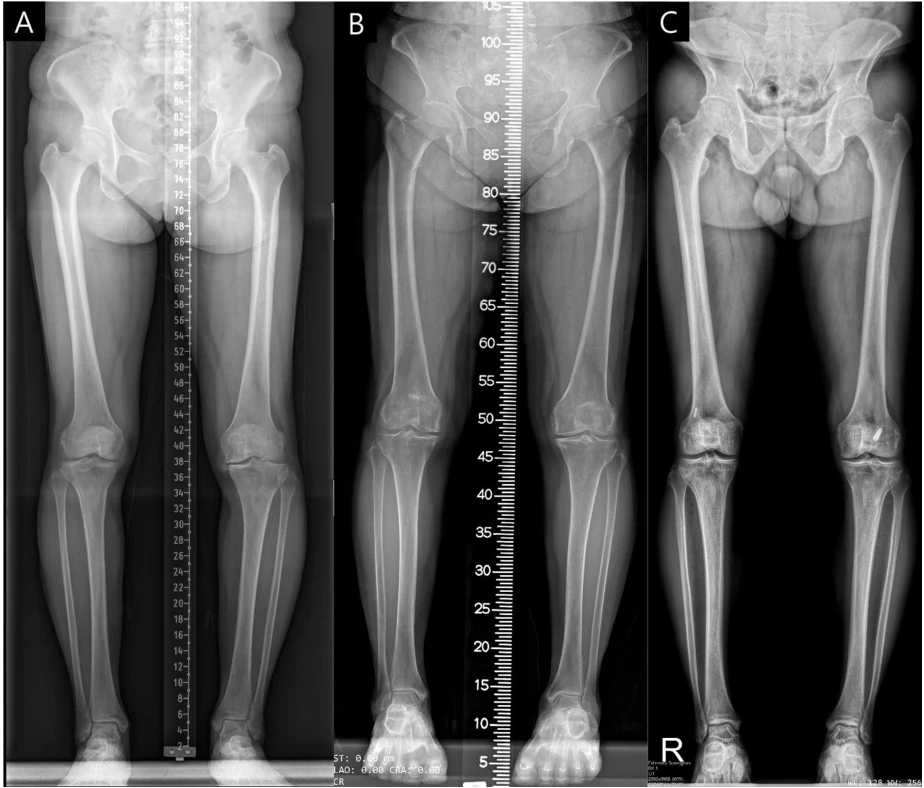
Original Figure 2 · X-rays from three institutions
 Enlarge Figure
Images from the three external-validation sites (A, B and C), used to assess landmark recognition across differences in contrast and field of view.
Sung Eun Kim, Jun Woo Nam, Joong Il Kim, Jong-Keun Kim, Du Hyun Ro. Knee Surgery & Related Research (2024). DOI: 10.1186/s43019-023-00209-y
The research question
Participants and Methods
Main Findings
Implications for clinical and surgical practice
Limitations
Let's put it briefly.
Which clinical question does this address?
This external validation relates to CONNEVO Metric measurement technology. Rapid repeat measurements should be accompanied by checking landmarks on the original image and considering positioning and intra-articular deformity.
Keywords
- Accuracy
- Alignment measurement
- Deep learning
- Full-leg radiographs
- Imaging protocol
- AI
- Metric
- alignment
Abstract · Original text
Sung Eun Kim, Jun Woo Nam, Joong Il Kim, Jong-Keun Kim, Du Hyun Ro. Enhanced deep learning model enables accurate alignment measurement across diverse institutional imaging protocols. Knee surgery & related research. 2024;36(1):4. doi:10.1186/s43019-023-00209-y.
BACKGROUND
Achieving consistent accuracy in radiographic measurements across different equipment and protocols is challenging. This study evaluates an advanced deep learning (DL) model, building upon a precursor, for its proficiency in generating uniform and precise alignment measurements in full-leg radiographs irrespective of institutional imaging differences.
METHODS
The enhanced DL model was trained on over 10,000 radiographs. Utilizing a segmented approach, it separately identified and evaluated regions of interest (ROIs) for the hip, knee, and ankle, subsequently integrating these regions. For external validation, 300 datasets from three distinct institutes with varied imaging protocols and equipment were employed. The study measured seven radiologic parameters: hip-knee-ankle angle, lateral distal femoral angle, medial proximal tibial angle, joint line convergence angle, weight-bearing line ratio, joint line obliquity angle, and lateral distal tibial angle. Measurements by the model were compared with an orthopedic specialist's evaluations using inter-observer and intra-observer intraclass correlation coefficients (ICCs). Additionally, the absolute error percentage in alignment measurements was assessed, and the processing duration for radiograph evaluation was recorded.
RESULTS
The DL model exhibited excellent performance, achieving an inter-observer ICC between 0.936 and 0.997, on par with an orthopedic specialist, and an intra-observer ICC of 1.000. The model's consistency was robust across different institutional imaging protocols. Its accuracy was particularly notable in measuring the hip-knee-ankle angle, with no instances of absolute error exceeding 1.5 degrees. The enhanced model significantly improved processing speed, reducing the time by 30-fold from an initial 10-11 s to 300 ms.
CONCLUSIONS
The enhanced DL model demonstrated its ability for accurate, rapid alignment measurements in full-leg radiographs, regardless of protocol variations, signifying its potential for broad clinical and research applicability.
© The Author(s) 2024
The original paper.
Enhanced deep learning model enables accurate alignment measurement across diverse institutional imaging protocols
Sung Eun Kim, Jun Woo Nam, Joong Il Kim, Jong-Keun Kim, Du Hyun Ro
Knee Surgery & Related Research · 2024