2025 · Journal of Clinical Medicine
Can lumbosacral transitional vertebrae be detected automatically on lateral X-rays?
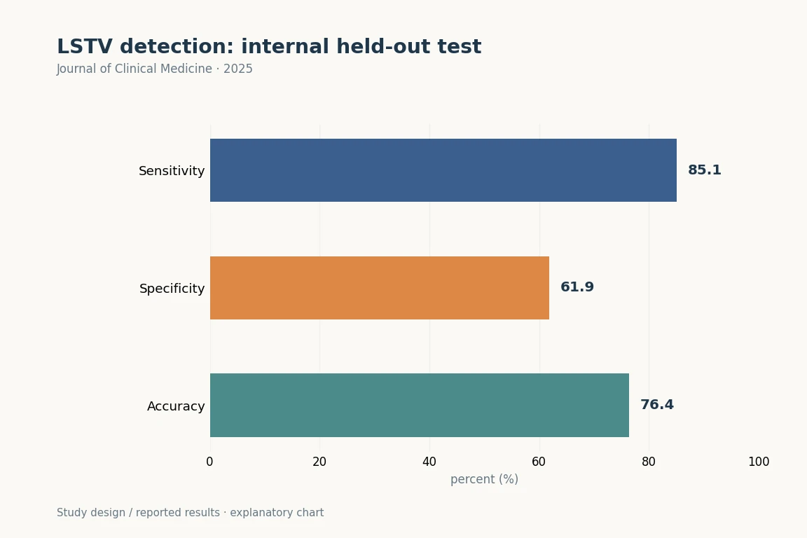
Test AUC was 0.84, sensitivity 85.1%, specificity 61.9% and accuracy 76.4%.
Results explained with study numbers
 Enlarge Figure
In the held-out test set of 313 images, sensitivity was 85.1%, specificity 61.9% and accuracy 76.4%.
Journal of Clinical Medicine (2025). DOI: 10.3390/jcm14217671 · Original explanatory chart based on reported data
The research question
Participants and Methods
Main Findings
Implications for clinical and surgical practice
Limitations
Let's put it briefly.
Which clinical question does this address?
This study supports identifying vertebral segments before spine surgery. It is a detection task separate from SPIN angle measurement.
Keywords
- artificial intelligence
- automated detection
- convolutional neural network
- deep learning
- lumbosacral transitional vertebrae
- plain radiograph
- spine imaging
- AI
Abstract · Original text
Donghyuk Kwak, Du Hyun Ro, Dong-Ho Kang. Automated Detection of Lumbosacral Transitional Vertebrae on Plain Lumbar Radiographs Using a Deep Learning Model. Journal of clinical medicine. 2025;14(21):7671. doi:10.3390/jcm14217671.
Background/Objectives: Lumbosacral transitional vertebra (LSTV) is a common anatomical variant, but its identification on plain radiographs is often inconsistent. This inconsistency can lead to clinical complications such as chronic low back pain, misinterpretation of spinal parameters, and an increased risk of wrong-level surgery. This study aimed to develop and validate a deep learning-based artificial intelligence (AI) model for the automated detection of LSTV on plain lumbar radiographs. Methods: This retrospective observational study included a total of 3116 standing lumbar lateral radiographs. The presence or absence of lumbosacral transitional vertebra (LSTV) was definitively established using whole-spine imaging, CT, or MRI. Multiple deep learning architectures, including DINOv2, CLIP (ViT-B/32), and ResNet-50, were initially evaluated for binary classification of LSTV. Among these, the ResNet-50 model with partial fine-tuning achieved the best test performance and was subsequently selected for fivefold cross-validation using the training set. Model performance was assessed using accuracy, sensitivity, specificity, and the area under the receiver operating characteristic curve (AUROC), and interpretability was evaluated using gradient-weighted class activation mapping (Grad-CAM). Results: On the independent test set of 313 radiographs, the final model demonstrated robust diagnostic performance. It achieved an accuracy of 76.4%, a sensitivity of 85.1%, a specificity of 61.9%, and an AUC of 0.84. The model correctly identified 166 out of 195 LSTV cases and 73 out of 118 normal cases. Conclusions: This AI-based system offers a highly accurate and reliable method for the automated detection of LSTV on plain radiographs. It shows strong potential as a clinical decision-support tool to reduce diagnostic errors, improve pre-operative planning, and enhance patient safety.
© 2025 by the authors.
The original paper.
Automated Detection of Lumbosacral Transitional Vertebrae on Plain Lumbar Radiographs Using a Deep Learning Model
Donghyuk Kwak, Du Hyun Ro, Dong-Ho Kang
Journal of Clinical Medicine · 2025