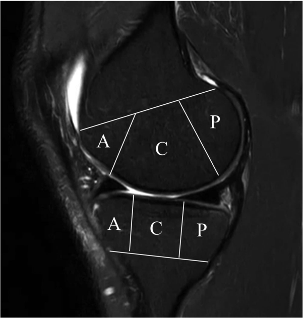
The X-ray intervals also contain the effects of cartilage.
An X-ray may show narrowing between the bones, but joint space is not a direct measure of cartilage thickness alone. Meniscal position and damage, and imaging posture, also matter. MRI distinguishes cartilage, meniscal, and bone-marrow lesions in greater detail. This study examined how information from the two tests relates over time.
So we divided the same knee into locations.
The study analyzed 3,710 knees over 7 years in MOST. Central and posterior MRI regions were assessed for cartilage damage, meniscal damage/extrusion and bone changes against X-ray grades. A, C and P mean anterior, central and posterior, distinguishing patterns that can differ within one knee.
We've seen different patterns in the center and back.
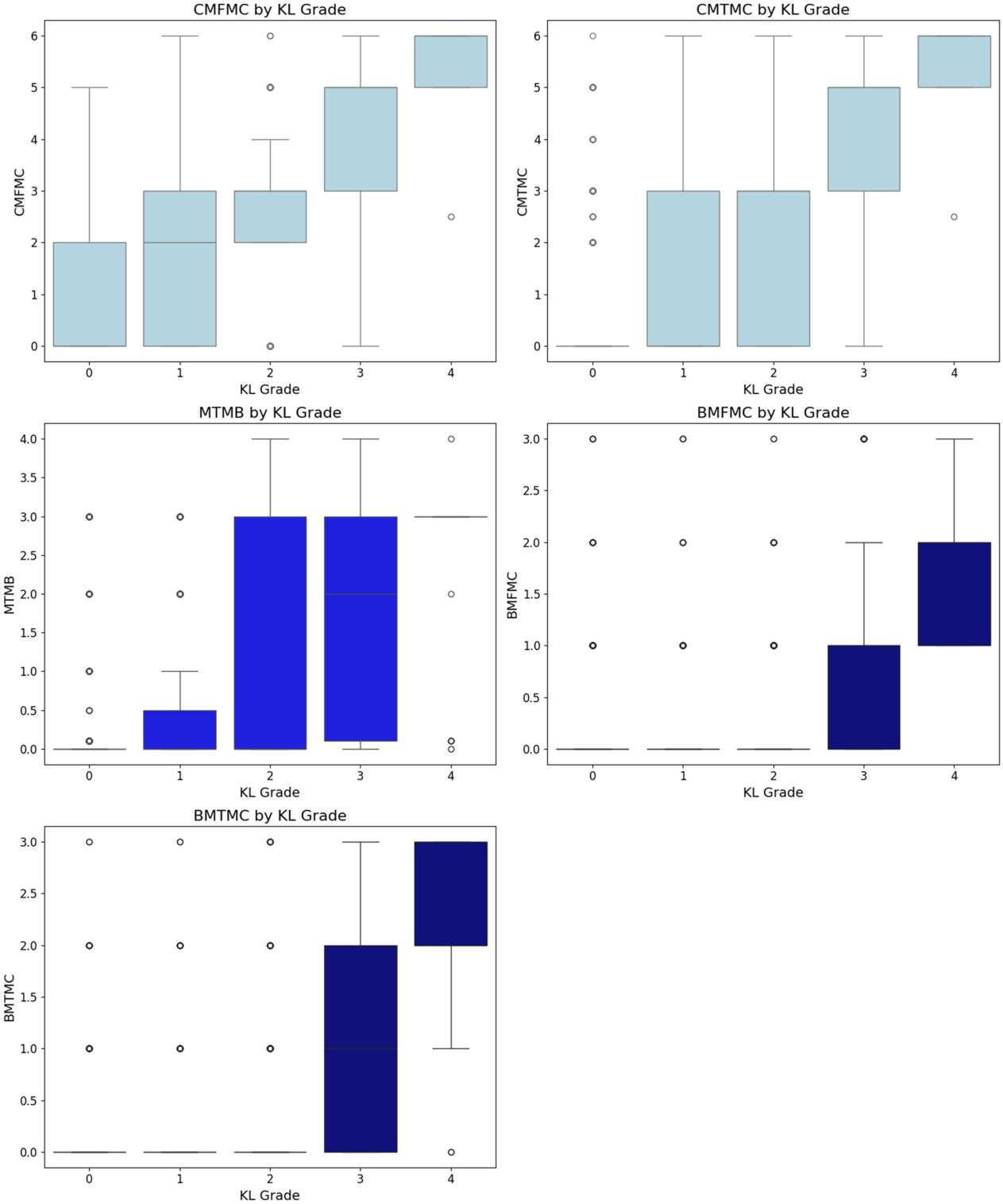
Cartilage changes appeared first centrally, while meniscal changes appeared first posteriorly. Tibial osteophytes were relatively early X-ray findings. Boxes and points show group distributions, not an identical sequence or precise timetable for every patient.
Original Figure 2
 Enlarge Figure
MRI findings of cartilage, meniscus and bone marrow in the central compartment are compared by X-ray KL grade.
Do Weon Lee, Ji-Sahn Kim, Hyuk-Soo Han, Du Hyun Ro. Longitudinal correlation between X-ray and MRI findings in medial compartment knee osteoarthritis: Insights into early cartilage loss and structural changes. Knee Surgery, Sports Traumatology, Arthroscopy. 2026;34(6):2047–2056. https://doi.org/10.1002/ksa.70016. © 2025 The Author(s).
Original Figure Legend
Figure 2
Meniscal extrusion emerged as an important finding
Meniscal extrusion beyond the joint margin had the strongest association among the examined indicators of later progression. Cartilage damage and meniscal tears were also associated. This does not prove that treating extrusion alone stops arthritis. The imaging associations provide a basis for further research and clinical evaluation.
Do you have to take an MRI for arthritis?
These findings do not recommend routine MRI for every patient. Symptoms, examination, and X-rays when needed remain important in OA assessment. Additional testing should address a specific clinical question, such as unexplained symptoms or suspected associated injury. Research seeks detailed tissue information; clinical care first asks what question a test will answer.