2025 · The Spine Journal
Can Pelvic and Lumbar Angles Be Measured Automatically on Radiographs of a Laterally Curved Spine?
Repeat-measurement ICC was 1.00 for every angle. Agreement with reference measurements and cross-validation ICCs were 0.96–0.99.
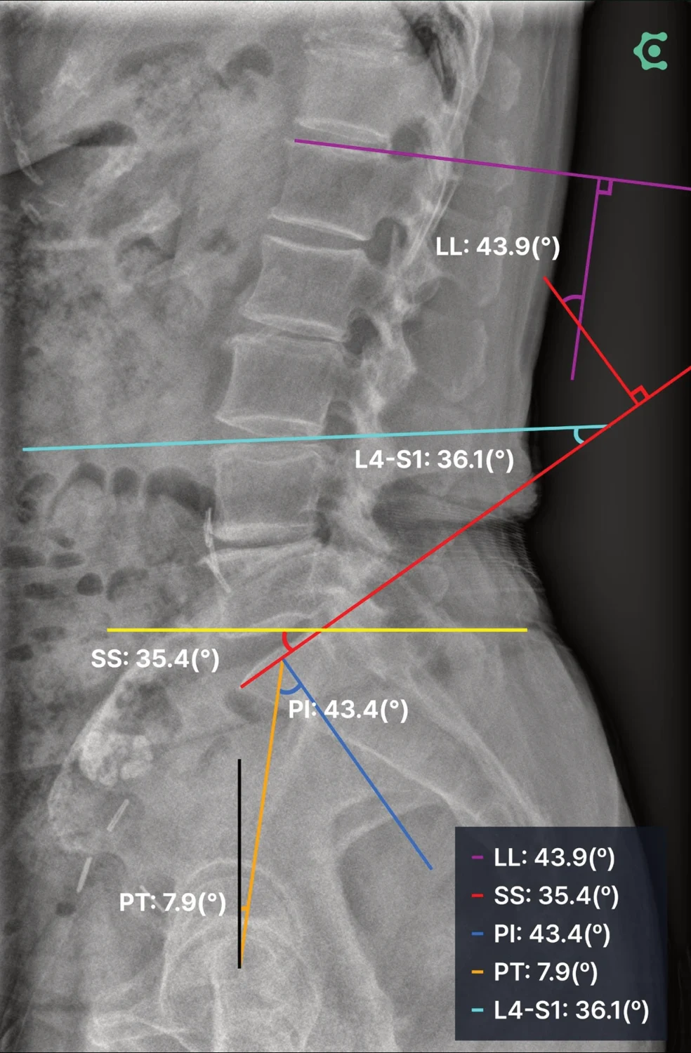
Lateral X-ray illustrating spinopelvic angles
 Enlarge Figure
A lateral X-ray showing LL, PI, PT, SS and the reference landmarks.
CONNECTEVE · CONNEVO SPIN · Owner-supplied product brochure
Results explained with study numbers
 Enlarge Figure
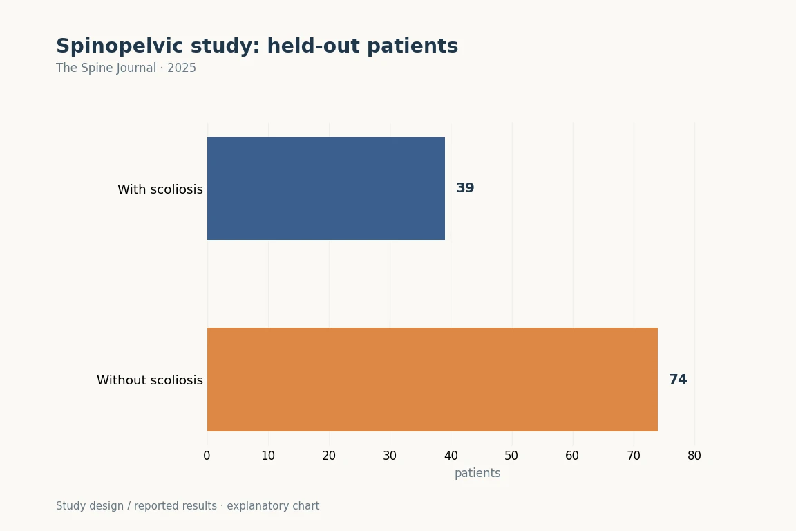
Of 113 test patients, 39 had degenerative scoliosis. LL, PI, PT and SS measurement differs from diagnostic classification. Repeat ICC 1.00 for the same image must be distinguished from agreement with specialist reference values.
The Spine Journal (2025). DOI: 10.1016/j.spinee.2025.01.020 · Original explanatory chart based on reported data
The research question
Participants and Methods
Main Findings
Implications for clinical and surgical practice
Limitations
Let's put it briefly.
Which clinical question does this address?
Automatic measurement may reduce the burden of repeatedly measuring spinopelvic alignment. When interpreting PI–LL mismatch and compensatory PT, first check image quality, femoral-head detection, and errors in the individual image.
Keywords
- Artificial intelligence
- Convolutional neural network
- Coronal deformity
- Keypoint detection
- Pelvic parameter
- Sagittal alignment
- Segmentation
- Neural Networks, Computer
Abstract · Original text
Dong-Ho Kang, Ye-Jin Jeong, Sung Taeck Kim, Younguk Kim, Bong-Soon Chang, Hyoungmin Kim, Sam Yeol Chang, Du Hyun Ro. Automated measurement of pelvic parameters using convolutional neural network in complex spinal deformities: overcoming challenges in coronal deformity cases. The spine journal : official journal of the North American Spine Society. 2025;25(8):1688-1697. doi:10.1016/j.spinee.2025.01.020.
BACKGROUND CONTEXT
Accurate and consistent measurement of sagittal alignment is challenging, particularly in patients with severe coronal deformities, including degenerative lumbar scoliosis (DLS).
PURPOSE
This study aimed to develop and validate an artificial intelligence (AI)-based system for automating the measurement of key sagittal parameters, including lumbar lordosis, pelvic incidence, pelvic tilt, and sacral slope, with a focus on its applicability across a wide range of deformities, including severe coronal deformities, such as DLS.
DESIGN
Retrospective observational study.
PATIENT SAMPLE
A total of 1,011 standing lumbar lateral radiographs, including DLS.
OUTCOME MEASURE
Interclass and intraclass correlation coefficients (CC), and Bland-Altman plots.
METHODS
The model utilizes a deep-learning framework, incorporating a U-Net for segmentation and a Keypoint Region-based Convolutional Neural Network for keypoint detection. The ground truth masks were annotated by an experienced orthopedic specialist. The performance of the model was evaluated against ground truth measurements and assessments from two expert raters using interclass and intraclass CC, and Bland-Altman plots.
RESULTS
In the test set of 113 patients, 39 (34.5%) had DLS, with a mean Cobb's angle of 14.8°±4.4°. The AI model achieved an intraclass CC of 1.00 across all parameters, indicating perfect consistency. Interclass CCs comparing the AI model to ground truth ranged from 0.96 to 0.99, outperforming experienced orthopedic surgeons. Bland-Altman analysis revealed no significant systemic bias, with most differences falling within clinically acceptable ranges. A 5-fold cross-validation further demonstrated robust performance, with interclass CCs ranging from 0.96 to 0.99 across diverse subsets.
CONCLUSION
This AI-based system offers a reliable and efficient automated measurement of sagittal parameters in spinal deformities, including severe coronal deformities. The superior performance of the model compared with that of expert raters highlights its potential for clinical applications.
The original paper.
Automated measurement of pelvic parameters using convolutional neural network in complex spinal deformities: overcoming challenges in coronal deformity cases
Kang DH, Jeong YJ, Kim ST, Kim Y, Chang BS, Kim H, Chang SY, Ro DH
The Spine Journal · 2025