2023 · KSSTA
Can Detecting 15 Landmarks on Full-length Lower-limb X-rays Reproduce Alignment Angles Automatically?
Angle-specific inter-observer ICCs for the automatic model were ≥0.98; repeat-measurement ICC was 1.00.
Results explained with study numbers
 Enlarge Figure
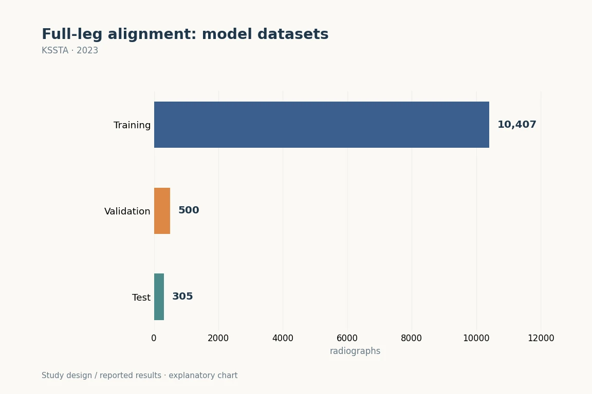
This single-institution study used 10,407 training, 500 validation and 305 test images. The figure shows the data split. Three-institution external validation was performed separately in the 2024 follow-up study.
KSSTA (2023). DOI: 10.1007/s00167-022-07124-x · Original explanatory chart based on reported data
The research question
Participants and Methods
Main Findings
Implications for clinical and surgical practice
Limitations
Let's put it briefly.
Which clinical question does this address?
The study supports standardizing mLDFA, MPTA, and HKAA measurements used at the start of HTO and TKA planning. Automatic measurement does not replace standardized imaging, interpretation of intra-articular deformity, or the surgeon’s final plan.
Keywords
- Angle measurement
- Deep learning
- Hip knee ankle angle (HKAA)
- Joint line convergence angle (JLCA)
- Landmark recognition
- Lower extremity
- Mechanical lateral distal femoral angle (mLDFA)
- Medial proximal tibial angle (MPTA)
Abstract · Original text
Changwung Jo, Doohyun Hwang, Sunho Ko, Myung Ho Yang, Myung Chul Lee, Hyuk-Soo Han, Du Hyun Ro. Deep learning-based landmark recognition and angle measurement of full-leg plain radiographs can be adopted to assess lower extremity alignment. Knee surgery, sports traumatology, arthroscopy : official journal of the ESSKA. 2023;31(4):1388-1397. doi:10.1007/s00167-022-07124-x.
PURPOSE
Evaluating lower extremity alignment using full-leg plain radiographs is an essential step in diagnosis and treatment of patients with knee osteoarthritis. The study objective was to present a deep learning-based anatomical landmark recognition and angle measurement model, using full-leg radiographs, and validate its performance.
METHODS
A total of 11,212 full-leg plain radiographs were used to create the model. To train the data, 15 anatomical landmarks were marked by two orthopaedic surgeons. Mechanical lateral distal femoral angle (mLDFA), medial proximal tibial angle (MPTA), joint line convergence angle (JLCA), and hip-knee-ankle angle (HKAA) were then measured. For inter-observer reliability, the inter-observer intraclass correlation coefficient (ICC) was evaluated by comparing measurements from the model, surgeons, and students, to ground truth measurements annotated by an orthopaedic specialist with 14 years of experience. To evaluate test-retest reliability, all measurements were made twice by each measurer. Intra-observer ICCs were then derived. Performance evaluation metrics used in previous studies were also derived for direct comparison of the model's performance.
RESULTS
Inter-observer ICCs for all angles of the model were 0.98 or higher (p < 0.001). Intra-observer ICCs for all angles were 1.00, which was higher than that of the orthopaedic specialist (0.97-1.00). Measurements made by the model showed no significant systemic variation. Except for JLCA, angles were precisely measured with absolute error averages under 0.52 degrees and proportion of outliers under 4.26%.
CONCLUSIONS
The deep learning model is capable of evaluating lower extremity alignment with performance as accurate as an orthopaedic specialist with 14 years of experience.
LEVEL OF EVIDENCE
III, retrospective cohort study.
The original paper.
Deep learning-based landmark recognition and angle measurement of full-leg plain radiographs can be adopted to assess lower extremity alignment
Jo C, Hwang D, Ko S, Yang MH, Lee MC, Han HS, Ro DH
KSSTA · 2023 · online 2022