Hearing “grade 2” or “grade 4” osteoarthritis can make it sound as though a number decides your treatment. Doctors commonly use the Kellgren–Lawrence (KL) classification to summarize structural changes on knee X-rays from 0 to 4. The grade is a useful starting point, but it does not describe all your symptoms or your future course. Our research began with the questions that come after that number.
What does the KL grade assess on an X-ray?
The grade considers joint-space narrowing, bony spurs at the joint margins, increased density of the bone beneath cartilage, and changes in the shape of the bone ends. Joint-space narrowing indirectly reflects changes in cartilage and the meniscus; cartilage itself is not directly visible on an X-ray.
The table below explains the commonly used original KL criteria. Grade 1 describes doubtful changes, while definite osteophytes are seen from grade 2. Grade 4 describes marked narrowing and advanced bony changes. These numbers are not percentages of remaining cartilage or scores for pain.
| Grade | Main radiographic findings |
|---|---|
| KL 0 | No definite signs of osteoarthritis. |
| KL 1 | Doubtful joint-space narrowing and possible osteophytes. |
| KL 2 | Definite osteophytes; joint-space narrowing may be present. |
| KL 3 | Multiple osteophytes, definite joint-space narrowing and some sclerosis; possible bone-end deformity. |
| KL 4 | Large osteophytes, marked joint-space narrowing, severe sclerosis and definite bone-end deformity. |
Does KL grade 4 mean I need a knee replacement now?
Grade 4 means that the X-ray shows advanced osteoarthritis. Yet people with similar images may differ greatly in walking, sleep and daily activities. Surgery timing considers those limitations, examination findings, the effects of exercise and medication, and overall health.
A lower grade should not be used to dismiss pain. A meniscal injury, synovial inflammation or a problem elsewhere can contribute to symptoms. It helps to discuss what the image shows and what you experience separately, then bring both into treatment decisions.
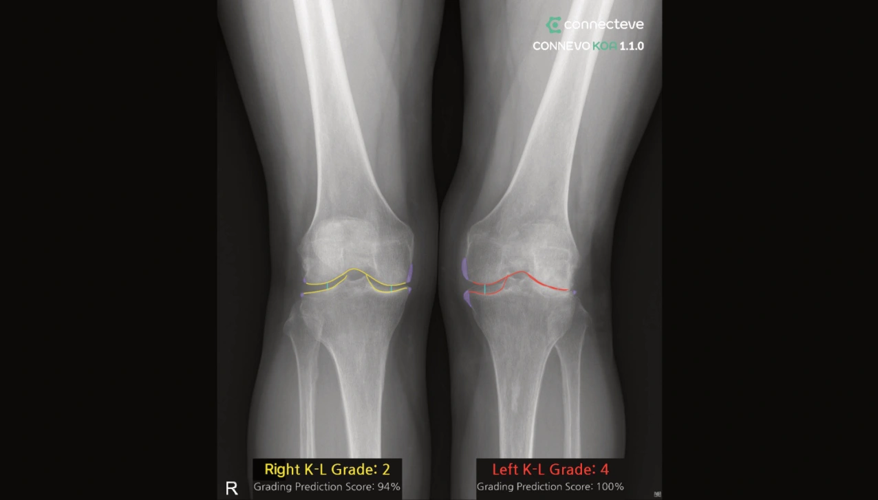
Knees with the same KL grade 2 can look and progress differently
Our group studied 10,804 knees from OAI and MOST, distinguishing knees dominated by osteophytes from those dominated by joint-space narrowing. Within the same early KL grade, pain and function did not differ significantly between these patterns, but progression to KL grade 3 or higher did.
Among KL grade 2 knees, the estimated mean time to progression to grade 3 or higher was about 5.4 times longer in the osteophyte-dominant group after adjustment for age, sex and BMI. This is not an individual countdown or a 5.4-fold difference in surgery risk. It shows why we ask which structural changes produced a grade, as well as what the grade is.
What are OAI and MOST?
OAI enrolled 4,796 men and women aged 45–79 in a long-term US osteoarthritis study. MOST is a separate longitudinal study that began with 3,026 people aged 50–79 and later added a new cohort. Repeated X-rays, MRIs and information on symptoms, function, weight and activity allow researchers to study changes in the same knees over time.
The number analyzed varies with available visits, variables and exclusion criteria. People and knees are also different counts. Testing a prediction method in an independent dataset helps check whether its performance extends beyond the population in which it was developed.
How did we study prediction of osteoarthritis progression?
In a separate study of knees initially graded KL 0–2, we developed a model using 5,966 OAI knees and externally validated it in 3,392 MOST knees. It predicted radiographic progression over 4–5 years using baseline KL grade, osteoarthritis in the opposite knee, medial and lateral narrowing, BMI and symptom/function questionnaire scores.
External-validation AUC was 0.76. AUC describes discrimination between knees that progressed and those that did not; it does not mean that a particular patient has a 76% probability of progression. The outcome was change in radiographic grade, rather than worsening pain or the date of knee replacement.
What should we discuss after the grade?
Ask which compartment is narrowed and what the osteophytes and leg alignment show. When comparing earlier images, check weight-bearing, knee bending and rotation. Differences caused by positioning should not automatically be read as disease progression.
Tell us what has changed in walking distance, stairs, sleep and exercise. KL grades summarize current structure, while detailed imaging and follow-up help us understand its course. Treatment is then matched to symptoms and personal goals. This is how more precise imaging research should return to patient care.