R.Du Hyun RoOrthopedics & Research
Text
Patient resourcesDisease and Treatment

Disease and Treatment

How does the meniscus distribute load? Understanding hoop stress

Circumferential fibers and root attachments work together to distribute load. The direction of a tear matters.

At a glance

  • No. It is a mechanical term for circumferential tensile stress during meniscal load transmission.
  • Repair and alternatives are considered with arthritis, alignment, tissue quality, symptoms and activity goals.

The meniscus is often called a shock-absorbing cushion, but compression alone does not explain how it works. To resist being pushed outward and spread load over a wider area, fibers running around its circumference must work with its attachments to bone. This connection helps explain why preserving the meniscus matters.

Compression creates tension around the circumference

The meniscus has a wedge-shaped cross-section. Compression from the femur tends to push it outward. Circumferential collagen fibers resist that displacement in tension, while the anterior and posterior roots maintain the attachment to bone. Circumferential tensile stress is called hoop stress; the associated tensile force is hoop tension.

This mechanism increases the area over which force is transmitted between the femur and tibia. Distributing the same force over a larger area reduces pressure at individual points. The continuity of fibers and roots matters alongside the amount of tissue.

Circumferential fibers and roots share the load

Connected fibers and roots
Posterior root detached
  • Force tending to push outward
  • Circumferential tension
  • Root attachment to bone
Conceptual top view of a meniscus. Arrows simplify force directions; the drawing does not reproduce actual anatomy, pressure values or tear size.

Why the direction of a tear matters

A radial tear cuts across load-bearing circumferential fibers. A root tear disrupts the attachment that holds the meniscus in place. Function can be substantially impaired even when much of the tissue remains. Longitudinal tears and horizontal splits have different geometry, so tear size or the word “tear” alone does not determine treatment.
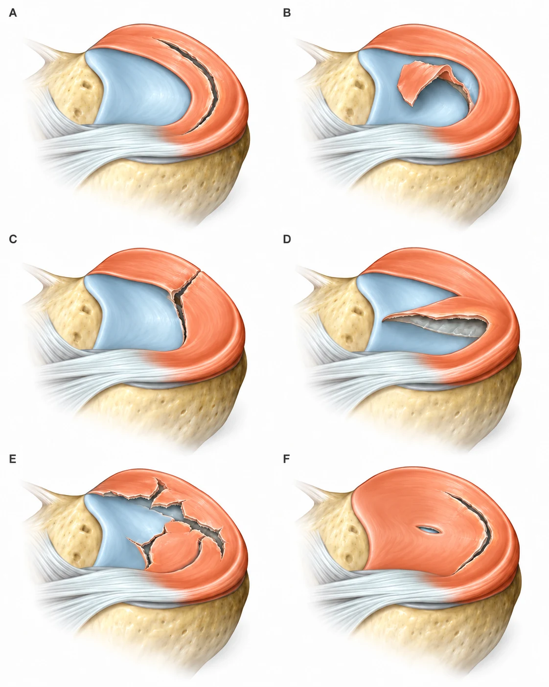
Patterns of meniscal tears
Tears have different directions and shapes. Symptoms and associated arthritis matter alongside the tear pattern. Orange indicates the meniscus; blue indicates articular cartilage.

Why a root tear can have a large mechanical effect

In a biomechanical experiment, a medial meniscus posterior root tear increased peak medial contact pressure by 25%, similar to total medial meniscectomy under the same conditions. Repair restored pressure to the intact level. This experiment used nine cadaveric knees; it does not establish an identical pressure change or rate of arthritis in every patient.

The key point is that a small part of the meniscus can be an important link in load transmission. MRI assessment considers tear location, extrusion beyond the joint margin and the condition of articular cartilage, alongside leg alignment.
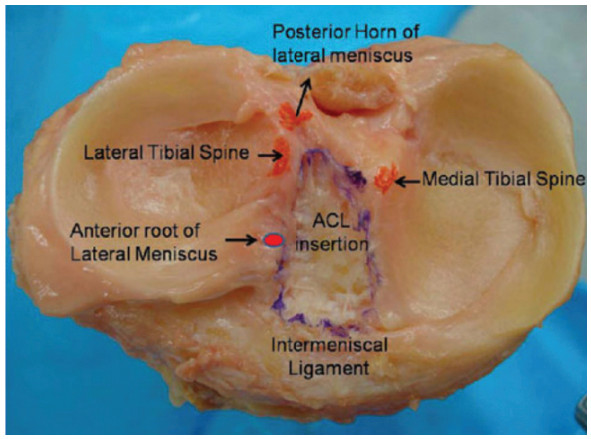
Original anatomical photograph shows meniscal roots and their relationship to the ACL insertion on the tibial plateau.

When are preservation and repair considered?

Preserving viable tissue and fiber continuity is an important goal. Repair suitability depends on tear pattern, blood supply, tissue quality and arthritis severity. Advanced arthritis or substantial malalignment may not be addressed by meniscal repair alone.

After repair, restrictions on loading and bending may be needed to allow healing. Ask which attachment or fiber continuity was restored and how it should be protected, as well as when walking can begin.
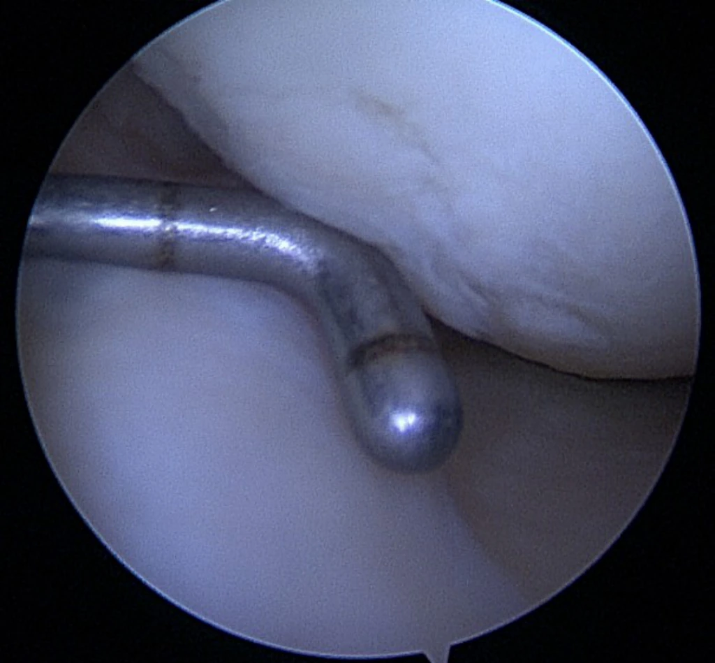
Arthroscopic view of a meniscal root examined with a probe
A probe is used to examine a meniscal root during arthroscopy. Tear location, tissue quality and attachment continuity are assessed. From the knee MRI teaching materials.
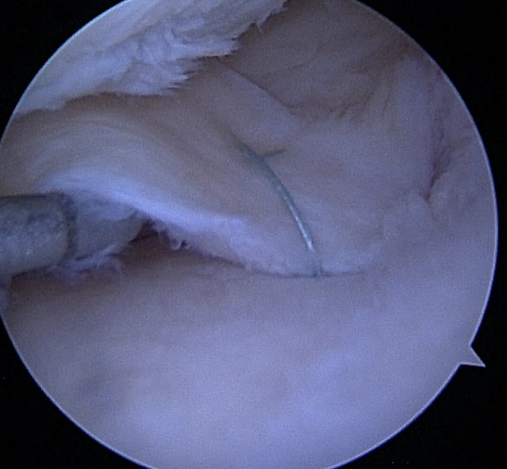
Arthroscopic view of the meniscal root region with a suture in place
A suture is visible in the meniscal root region. Repair suitability and postoperative protection and rehabilitation depend on the tear pattern, tissue and joint condition. From the knee MRI teaching materials.

What should I ask about my meniscus image?

Ask whether the tear crosses the circumferential fibers, whether the roots remain attached and whether extrusion is present. Discuss arthritis, repair options and the protection and rehabilitation needed afterward. Preservation means making choices that improve current symptoms while retaining useful tissue for the long term.

Common Questions

Is hoop stress a pain score?

No. It is a mechanical term for circumferential tensile stress during meniscal load transmission.

Does every root tear require repair?

Repair and alternatives are considered with arthritis, alignment, tissue quality, symptoms and activity goals.

Read more

Allaire et al. Biomechanical consequences of a medial meniscus posterior root tear. JBJS Am, 2008.AAOS OrthoInfo · Meniscus Tears

When to seek medical care

Each warning sign is a separate reason to seek care. Follow the action below if any one applies.

Emergency department · call 119 in Korea

  • Call 119 for sudden breathlessness or chest pain, severe faintness or loss of consciousness. Do not wait for calf swelling to appear.
  • Go to an emergency department if you suddenly cannot bear any weight, or if an injured knee swells rapidly or looks deformed. A cold or numb foot after injury also needs immediate assessment.
  • Seek emergency care for a hot, swollen and severely painful joint with high fever, shaking chills, confusion or inability to bear weight.
  • With back or leg pain, go immediately to an emergency department for new difficulty passing urine, loss of bladder or bowel control, or numbness around the genitals or anus. Severe or rapidly worsening weakness or numbness in both legs also needs immediate assessment.

Same-day assessment

  • Arrange assessment today for a newly hot, swollen joint, sudden severe pain, or a knee locked so that it cannot fully straighten. Joint infection can occur without fever.
  • Have new swelling or pain in one calf assessed today for a possible blood clot. Call 119 immediately if breathlessness or chest pain develops.
  • Severe knee pain or a new limp in a growing adolescent may come from a hip condition such as slipped capital femoral epiphysis. Stop walking and sport and arrange assessment today. Go directly to emergency care if they cannot bear weight.
  • Stop running and seek assessment today for new recurring groin pain, pain with walking or at night, or a limp. A femoral neck stress fracture needs to be considered. Keep weight off the painful leg until assessed. Go to emergency care if you cannot bear weight.

Book an outpatient appointment

  • If none of the warning signs above is present, book an appointment for pain lasting a few weeks, repeated swelling or reduced daily activity and exercise.