R.Du Hyun RoOrthopedics & Research
Text
← The Story of Research

arthritis inspection and surveillance · 2024

Where is the groove at the front of the knee shallower?

Dividing the patellar groove into 3-dimensional regions revealed shape differences, particularly proximally.

Knee Surgery, Sports Traumatology, Arthroscopy · Joon Young Lee, Sung Eun Kim, Oh Hyo Kwon, Youngjun Kim, Tae-Geun Son, Hyuk-Soo Han, Du Hyun Ro

Original Figure 2

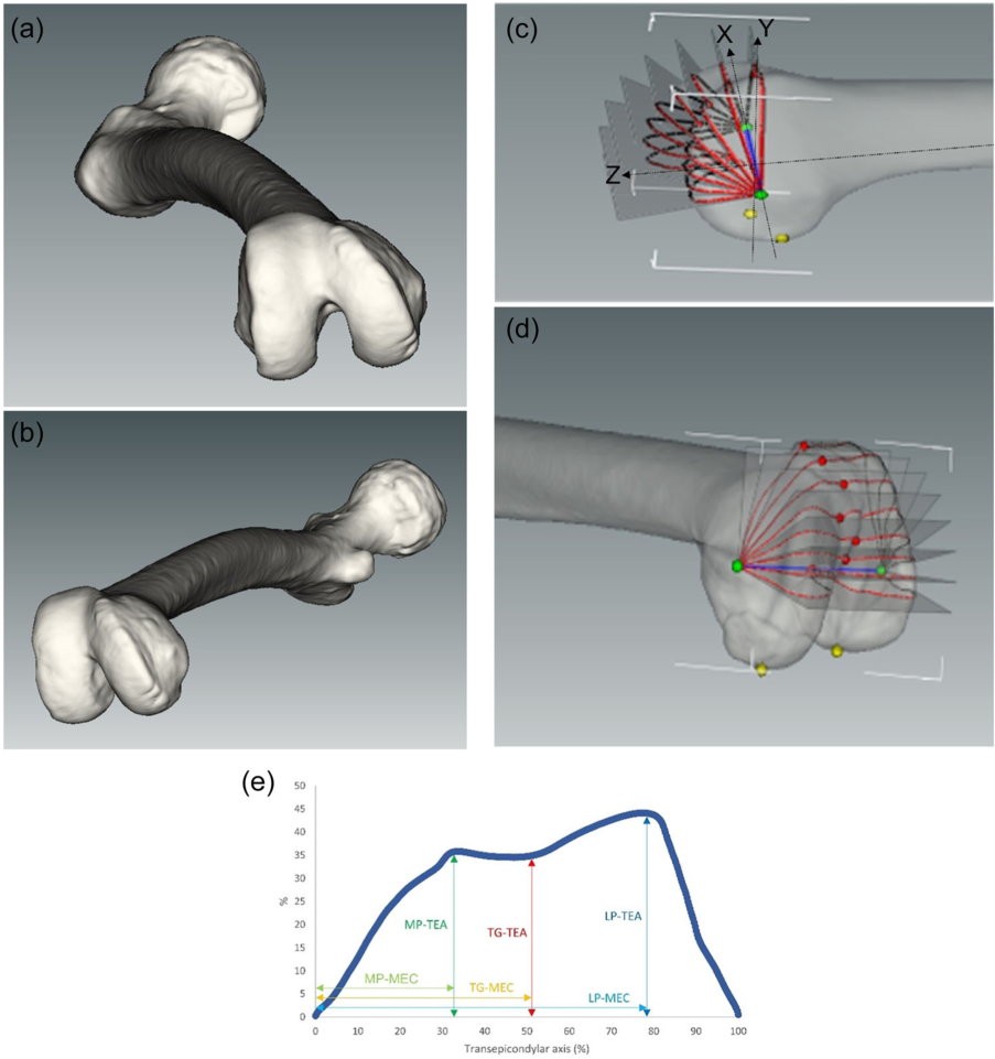
We use 3D CT to reconstruct the shape of the bow and take a steady axis and compare pieces at different heights. It's a way of describing where is narrower than the classification that you see in a side photo.Enlarge Figure

We use 3D CT to reconstruct the shape of the bow and take a steady axis and compare pieces at different heights. It's a way of describing where is narrower than the classification that you see in a side photo.

Joon Young Lee, Sung Eun Kim, Oh Hyo Kwon, Youngjun Kim, Tae-geun Son, Hyuk-Soo Han, Du Hyun Ro. Shallow trochlear groove and narrow medial trochlear width at the proximal trochlea in patients with trochlear dysplasia: A three-dimensional computed tomography analysis. Knee Surgery, Sports Traumatology, Arthroscopy. 2024;32(6):1434–1445. https://doi.org/10.1002/ksa.12166.

Evidence & Original SourceImage use terms
Original Figure Legend

Figure 2

01

I see the way patella moves.

The patella moves in the groove at the end of the femur, called the trochlea. A shallow or differently shaped groove can be associated with patellar instability, but not every case of anterior knee pain is trochlear dysplasia. This research describes trochlear shape more precisely rather than comparing pain treatments.

02

Instead of a single image, we compared several pieces of 3D CT.

3D CTs from 20 trochlear-dysplasia patients and 40 controls were reconstructed. A consistent bone axis defined 8 planes from 0–105° at 15° intervals. Standard measurements of groove depth and medial/lateral width reduce subjective classification differences and describe regional features.

03

The difference was particularly pronounced on the upper arches.

The dysplasia group had a shallower groove and narrower medial width in the proximal, or upper, trochlea. Differences were not uniform at every level, making slice position important. The models and axes show where each distance was measured and help explain the numerical results.

Original Figure 3
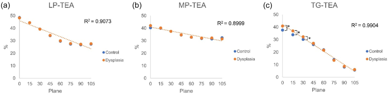
It's a basic diagram that compares how the gradient pattern changes when the angles of the intersection change. If the measurement position is different, then an indicator with the same name can produce a different value.Enlarge Figure

It's a basic diagram that compares how the gradient pattern changes when the angles of the intersection change. If the measurement position is different, then an indicator with the same name can produce a different value.

Joon Young Lee, Sung Eun Kim, Oh Hyo Kwon, Youngjun Kim, Tae-geun Son, Hyuk-Soo Han, Du Hyun Ro. Shallow trochlear groove and narrow medial trochlear width at the proximal trochlea in patients with trochlear dysplasia: A three-dimensional computed tomography analysis. Knee Surgery, Sports Traumatology, Arthroscopy. 2024;32(6):1434–1445. https://doi.org/10.1002/ksa.12166.

Original Figure Legend

Figure 3

04

It's a study that connects classification to actual forms.

The Dejour classification is an important clinical tool, but image quality and reader differences can affect judgments. Adding 3-dimensional quantitative measures allows research into regional differences within a type. Further validation is needed to show how well those measures explain postoperative redislocation risk or symptoms.

05

CT isn't necessary for every patient.

Use of CT in a study does not mean every person with anterior knee pain needs CT. Radiation and the diagnostic question matter. Examination and other images guide its use for recurrent dislocation or surgical planning. Treatment considers patellar position, ligaments and leg alignment, not trochlear shape alone.

Look at the study numbers again.

20Participants with trochlear dysplasia
40Comparison controls
8Cross-sections compared
RESEARCH FINDINGknee

Composition of the study sample

Participants with trochlear dysplasia
20
01223
Counts of data included in the study. These do not show treatment effects or an individual’s risk.

Common Questions

Can we apply these results directly to individual therapies?

Use of CT in a study does not mean every person with anterior knee pain needs CT. Radiation and the diagnostic question matter. Examination and other images guide its use for recurrent dislocation or surgical planning. Treatment considers patellar position, ligaments and leg alignment, not trochlear shape alone.

Methods and Numbers in Detail

Shallow trochlear groove and narrow medial trochlear width at the proximal trochlea in patients with trochlear dysplasia: A three-dimensional computed tomography analysis

Participants and Methods

20 people with Trochlear dysplasia and 40 health counterparts.

In 3D CT reconstructions, 6 TEA- and MEC-related distances were measured in 8 planes from 0–105°, at 15° intervals.

Results Reported in the Paper

  1. TG-TEA and MP-MEC were greater in the trochlear dysplasia group in the 0°, 15°, and 30° planes (all P < 0.05).
  2. A shallow trochlear groove and a narrow medial trochlear width were reported in the proximal trochlea at 0–30°.

Scope of Interpretation

This was a morphological study. It did not compare the effects of trochleoplasty, MPFL, or TTO techniques. With 20 patients with dysplasia, generalization across subtypes is limited.

Keywords

Abstract · Original text

Joon Young Lee, Sung Eun Kim, Oh Hyo Kwon, Youngjun Kim, Tae-Geun Son, Hyuk-Soo Han, Du Hyun Ro. Shallow trochlear groove and narrow medial trochlear width at the proximal trochlea in patients with trochlear dysplasia: A three-dimensional computed tomography analysis. Knee surgery, sports traumatology, arthroscopy : official journal of the ESSKA. 2024;32(6):1434-1445. doi:10.1002/ksa.12166.

PURPOSE

Although the Dejour classification is the primary classification system for evaluating trochlear dysplasia, concerns have been raised about its reliability owing to its qualitative criteria and challenges associated with obtaining accurate radiographs. This study aimed to quantify trochlear dysplasia using three-dimensional (3D) computed tomography (CT) reconstruction with novel parameters related to the transepicondylar axis (TEA).

METHODS

Sixty patients were enrolled, including 20 with trochlear dysplasia and 40 healthy controls. The 3D CT model was generated using the Materialise Interactive Medical Image Control System software. The following six parameters were measured in eight consecutive planes at 15° intervals (planes 0-105): the distance from the TEA to the most cortical point of the lateral condyle ('LP-TEA', where LP stands for lateral peak), medial condyle ('MP-TEA', MP for medial peak) and deepest point of the trochlea ('TG-TEA', TG for trochlear groove). The distances from the medial epicondyle (MEC) to the corresponding TEA points were measured ('LP-MEC', 'MP-MEC' and 'TG-MEC').

RESULTS

In the dysplasia group, TG-TEA (planes 0, 15 and 30) and MP-MEC (planes 0, 15 and 30) were significantly greater than those in the control group (all p < 0.05 for planes of TG-TEA and MP-MEC). For type A dysplasia, LP-MEC (plane 0) was greater than that in the control group. For type B dysplasia, the MP-MEC (planes 0 and 15) and TG-TEA (planes 0 and 15) were greater than those of the control group. For type D dysplasia, MP-MEC (planes 0, 15 and 30) and TG-TEA (planes 0 and 15) were elevated.

CONCLUSION

The 3D CT reconstruction analysis established a reproducible method for quantifying osseous trochlear morphology. Patients with trochlear dysplasia had a shallow TG and narrow medial trochlear width at tracking angles of 0°-30°. This finding corroborates the clinical manifestations of recurrent patellar instability that occur during early flexion.

LEVEL OF EVIDENCE

Level III.