Symptoms and tests
What tissues are inside the knee?
Understand the bones, cartilage, menisci, ligaments, muscles and synovial lining.
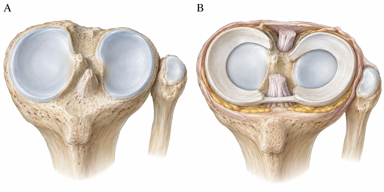
At a glance
- Articular cartilage and the menisci are different tissues.
- Stability depends on muscles and movement as well as ligaments.
- Describe both the location of pain and the activity that brings it on.
People may use the same words to describe knee pain even when the tissues involved and the reasons differ. Knowing the names mentioned during tests can help you understand the consultation and describe which movements are uncomfortable. Bone, ligaments, menisci and muscles work together in the knee, alongside cartilage.
Three roles within the knee
- 01
Joint surfaces
Cartilage and the bone beneath it carry load.
- 02
Load and stability
Menisci distribute load; ligaments limit excessive movement.
- 03
Movement and fluid
Muscles and tendons create movement; the synovial lining relates to joint fluid.
Three panels distinguish joint surfaces, stabilizing tissues and movement-related tissues
The joint surface and the bone beneath it
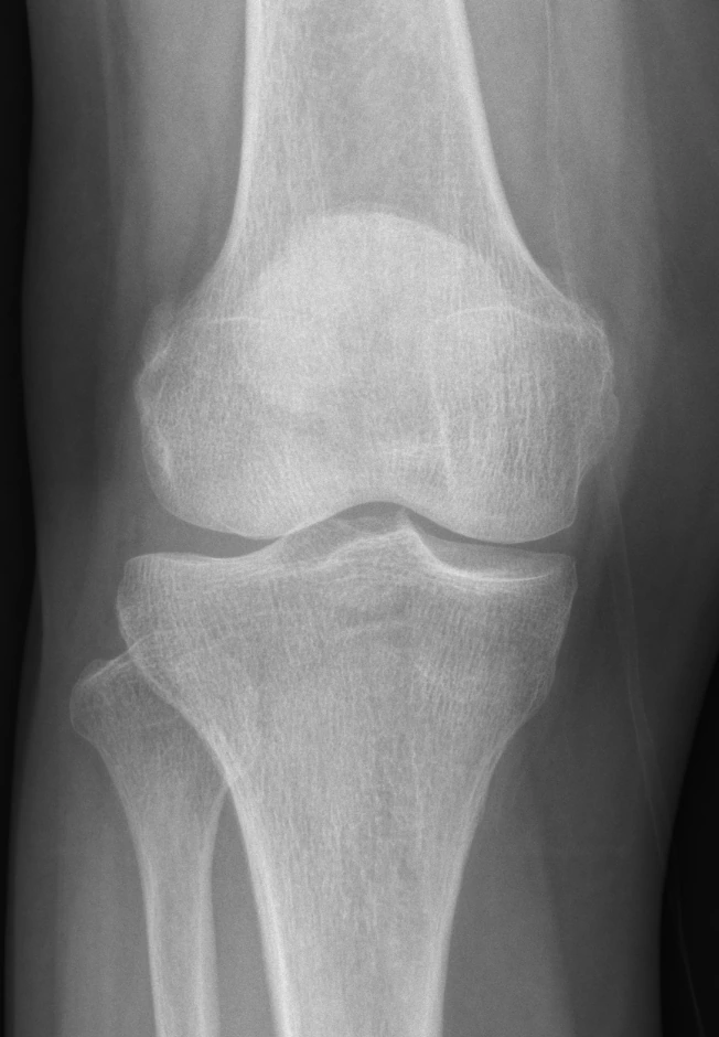
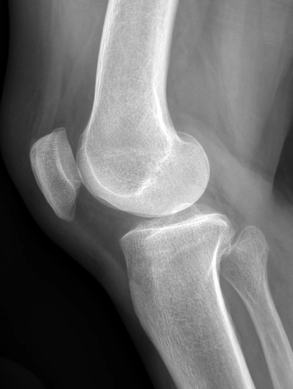
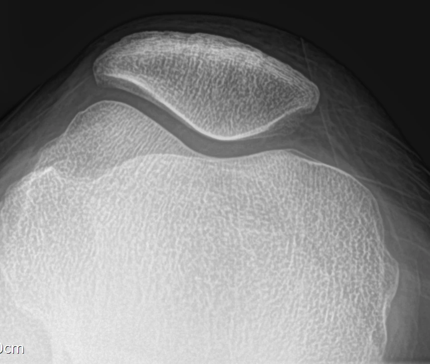
The thigh bone, or femur, meets the shin bone, or tibia, at the knee. The kneecap, or patella, also forms a joint with the femur. Articular cartilage covers the surfaces that meet and helps them move smoothly. The patellofemoral joint is considered when assessing pain at the front of the knee.
The bone under the cartilage also carries load. Osteoarthritis can involve this bone and bony spurs around the joint, as well as cartilage changes. An X-ray mainly shows bone and joint space. A description of narrowed joint space is therefore different from directly seeing the cartilage.
The menisci and ligaments
A meniscus is a crescent-shaped tissue between the femur and tibia. There is one on the inner side and one on the outer side. They distribute load and contribute to stability. Their position, structure and treatment after injury differ from those of articular cartilage.
The cruciate and collateral ligaments limit excessive movement between the bones. Buckling when turning and swelling after an injury are useful clues. However, a feeling of instability does not always mean a torn ligament: pain and muscle weakness can also play a part.
Muscles and the synovial lining also matter
The front thigh muscles, patella and tendons are linked in the mechanism that straightens the knee. The hamstrings and hip muscles contribute to walking and balance. Even when joint changes remain, better strength and movement can make walking or rising from a chair easier.
The synovial lining is related to the fluid inside the joint. Inflammation there can cause swelling or warmth. Tendons and bursae outside the joint can also hurt. Pain around the knee should not automatically be attributed to cartilage inside it.
Describe the location and the movement
Point to whether the pain is at the front, inner side, outer side or back. Also mention stairs, rising from a chair or changing direction if these bring it on. Location alone does not establish a diagnosis; examination findings are compared with imaging.
A consultation considers both the tissue involved and how much the problem limits daily life. This helps determine whether an imaging change actually explains the symptoms. Mention pain that changes location and any associated hip or back symptoms too.
Common Questions
Can an X-ray directly show cartilage?
Ordinary X-rays do not clearly show cartilage itself. Joint space and other findings help assess structural changes.
Does pain at the front mean a kneecap problem?
That is one possibility. Tendons, muscles and other structures must also be examined.
Does strengthening remove structural damage?
It does not reverse every change. Improving strength and function can still make everyday life easier.
Evidence and references
01When to seek medical care
Each warning sign is a separate reason to seek care. Follow the action below if any one applies.
Emergency department · call 119 in Korea
- Call 119 for sudden breathlessness or chest pain, severe faintness or loss of consciousness. Do not wait for calf swelling to appear.
- Go to an emergency department if you suddenly cannot bear any weight, or if an injured knee swells rapidly or looks deformed. A cold or numb foot after injury also needs immediate assessment.
- Seek emergency care for a hot, swollen and severely painful joint with high fever, shaking chills, confusion or inability to bear weight.
- With back or leg pain, go immediately to an emergency department for new difficulty passing urine, loss of bladder or bowel control, or numbness around the genitals or anus. Severe or rapidly worsening weakness or numbness in both legs also needs immediate assessment.
Same-day assessment
- Arrange assessment today for a newly hot, swollen joint, sudden severe pain, or a knee locked so that it cannot fully straighten. Joint infection can occur without fever.
- Have new swelling or pain in one calf assessed today for a possible blood clot. Call 119 immediately if breathlessness or chest pain develops.
- Severe knee pain or a new limp in a growing adolescent may come from a hip condition such as slipped capital femoral epiphysis. Stop walking and sport and arrange assessment today. Go directly to emergency care if they cannot bear weight.
- Stop running and seek assessment today for new recurring groin pain, pain with walking or at night, or a limp. A femoral neck stress fracture needs to be considered. Keep weight off the painful leg until assessed. Go to emergency care if you cannot bear weight.
Book an outpatient appointment
- If none of the warning signs above is present, book an appointment for pain lasting a few weeks, repeated swelling or reduced daily activity and exercise.