Symptoms and tests
How are cartilage and the meniscus different?
Similar names can hide different locations, roles and treatment choices.
At a glance
- Articular cartilage covers bone surfaces; menisci distribute load between bones.
- An MRI tear alone does not establish the cause of pain or the need for surgery.
- Clarify whether treatment aims at preservation, pain relief or improved function.
Hearing that cartilage is worn and a meniscus is torn can make them sound like the same tissue. They do different jobs inside the knee. Identifying which tissue has changed makes both the scan report and the treatment discussion easier to understand.
Two tissues with different roles
- 01
Articular cartilage
Tissue covering the joint surfaces at the ends of bones.
- 02
Meniscus
Crescent-shaped tissue distributing load between bones.
- 03
Treatment decision
Consider symptoms, the type of damage and remaining function together.
Three panels compare cartilage on bone surfaces, menisci between bones and treatment assessment
Articular cartilage covers the ends of bones
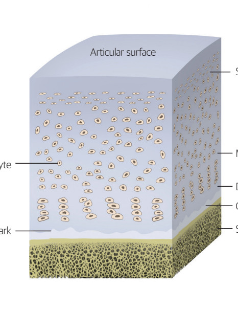
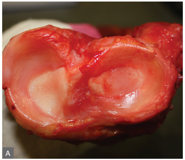
Articular cartilage is the smooth tissue covering the joint surfaces of the femur, tibia and patella. It helps reduce friction and carries load. Osteoarthritis may affect a broad area of the joint surface, while an injury can leave a more localized defect.
Widespread osteoarthritis and a small focal defect present different treatment conditions. The bone underneath, limb alignment, other joint surfaces, age and activity goals matter alongside defect size. An abnormality in cartilage does not by itself identify a suitable restoration procedure.
The menisci lie between the bones
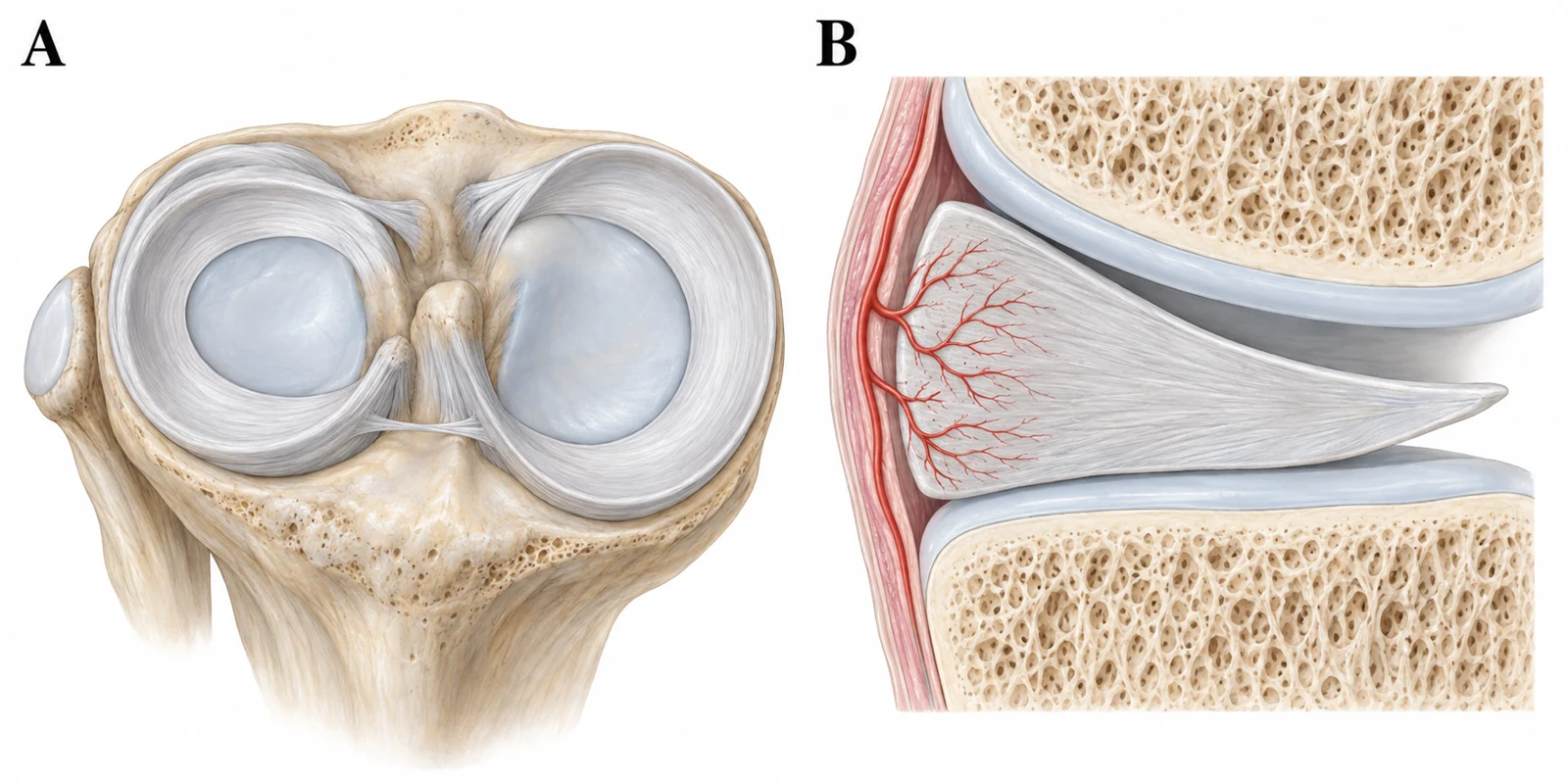
There is an inner and an outer meniscus between the femur and tibia. Their circumferential fibers and root attachments help distribute load. Treating them as unnecessary loose pieces overlooks the value of preserving functioning tissue.
Traumatic and degenerative tears are assessed differently. Shape, location, tissue quality, actual catching symptoms and associated arthritis matter. Seeing a tear on MRI is not the same as establishing that it is the main cause of the current pain.
Treatment considers function and preservation
Removing all the meniscus is not a route to a healthier knee. When surgery is appropriate, the aim is to preserve as much functioning tissue as possible and consider whether repair or limited removal is needed. Not every tear requires surgery; the response to nonsurgical treatment also matters.
For articular cartilage damage, discussions may include symptom control and strengthening, focal restoration, joint-preserving surgery or replacement, depending on the situation. The word cartilage in an injection name does not demonstrate that a worn surface has regrown. Ask what benefit is expected and how it will be assessed.
Three useful questions about a scan
Ask which tissue is affected and where, how the finding relates to your symptoms, and what the recommended treatment aims to improve. Rehabilitation after meniscal repair can differ from rehabilitation after partial removal, so it helps to know the exact procedure name.
A knee that suddenly locks and cannot straighten, or major swelling after an injury, deserves assessment rather than being dismissed as an old degenerative finding. Conversely, a scan abnormality with little discomfort and preserved function does not by itself create urgency for surgery.
Articular cartilage and the menisci protect the knee together
Articular cartilage provides a smooth covering over the ends of the bones to help the knee move. The medial and lateral menisci lie between the femur and tibia and are made of fibrocartilage. They transmit weight, distribute impact, and contribute to knee stability.
Fibres within each meniscus run around its circumference. Their continuity allows a downward compressive load to be carried as tension around the meniscus. Understanding this load-sharing function helps explain why the location and direction of a tear matter when considering treatment.
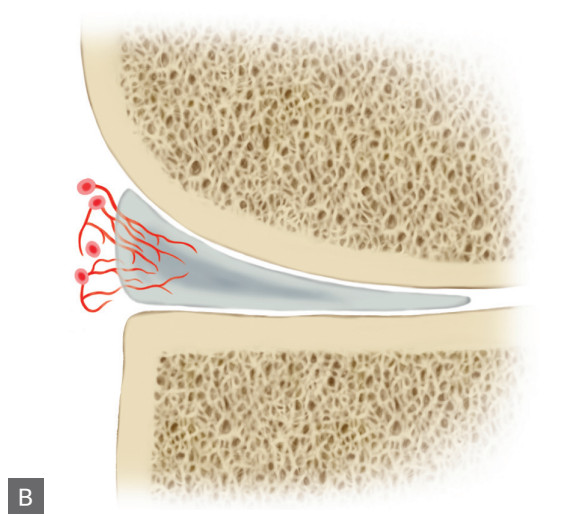
Within each meniscus, the edge near the joint capsule has a richer blood supply; blood supply and healing potential become more limited toward the centre. These regions are distinct from the medial and lateral menisci on the two sides of the knee. Treatment considers blood supply together with the tear pattern, tissue condition, and associated arthritis to assess healing potential and ways to preserve tissue.
Common Questions
Is the meniscus a type of cartilage?
It is fibrocartilage, but its structure and role differ from articular cartilage on the bone surface.
Can every meniscal tear be repaired?
Repairability depends on location, tear pattern, blood supply, tissue quality and associated problems.
Do abnormalities in both mean I need surgery?
Symptoms, examination, the extent of arthritis and the response to nonsurgical treatment must be considered together.
Evidence and references
01When to seek medical care
Each warning sign is a separate reason to seek care. Follow the action below if any one applies.
Emergency department · call 119 in Korea
- Call 119 for sudden breathlessness or chest pain, severe faintness or loss of consciousness. Do not wait for calf swelling to appear.
- Go to an emergency department if you suddenly cannot bear any weight, or if an injured knee swells rapidly or looks deformed. A cold or numb foot after injury also needs immediate assessment.
- Seek emergency care for a hot, swollen and severely painful joint with high fever, shaking chills, confusion or inability to bear weight.
- With back or leg pain, go immediately to an emergency department for new difficulty passing urine, loss of bladder or bowel control, or numbness around the genitals or anus. Severe or rapidly worsening weakness or numbness in both legs also needs immediate assessment.
Same-day assessment
- Arrange assessment today for a newly hot, swollen joint, sudden severe pain, or a knee locked so that it cannot fully straighten. Joint infection can occur without fever.
- Have new swelling or pain in one calf assessed today for a possible blood clot. Call 119 immediately if breathlessness or chest pain develops.
- Severe knee pain or a new limp in a growing adolescent may come from a hip condition such as slipped capital femoral epiphysis. Stop walking and sport and arrange assessment today. Go directly to emergency care if they cannot bear weight.
- Stop running and seek assessment today for new recurring groin pain, pain with walking or at night, or a limp. A femoral neck stress fracture needs to be considered. Keep weight off the painful leg until assessed. Go to emergency care if you cannot bear weight.
Book an outpatient appointment
- If none of the warning signs above is present, book an appointment for pain lasting a few weeks, repeated swelling or reduced daily activity and exercise.