
2026 · Knee Surgery, Sports Traumatology, Arthroscopy
How Are Radiographic OA Changes Related Over Time to Cartilage, Meniscal, and Bone Changes on MRI?
Centrally, meniscal and bone changes followed cartilage damage; posteriorly, meniscal changes appeared first. Central femoral cartilage loss was observed even at KL 0.
Original Figure 1
 Enlarge Figure
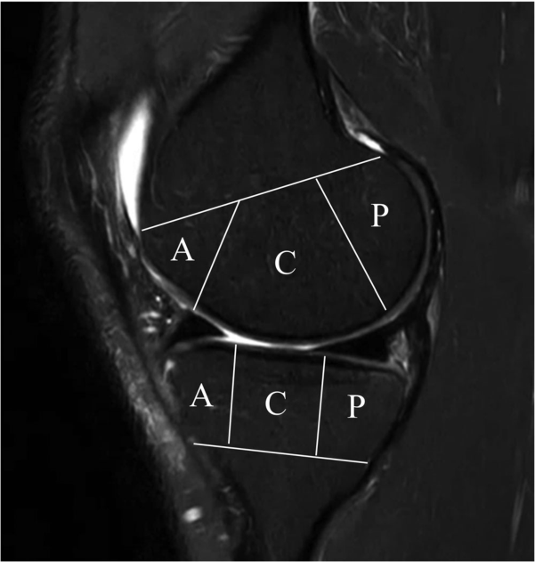
In an MRI, you divide the cartilage and cranial bone in front of A; in the middle of C; behind P. The same knee can vary the amount of tissue change depending on location, so we've broken down the layout.
Do Weon Lee, Ji-Sahn Kim, Hyuk-Soo Han, Du Hyun Ro. Longitudinal correlation between X-ray and MRI findings in medial compartment knee osteoarthritis: Insights into early cartilage loss and structural changes. Knee Surgery, Sports Traumatology, Arthroscopy. 2026;34(6):2047–2056. https://doi.org/10.1002/ksa.70016. © 2025 The Author(s).
Original Figure Legend
Figure 1
Original Figure 2
 Enlarge Figure
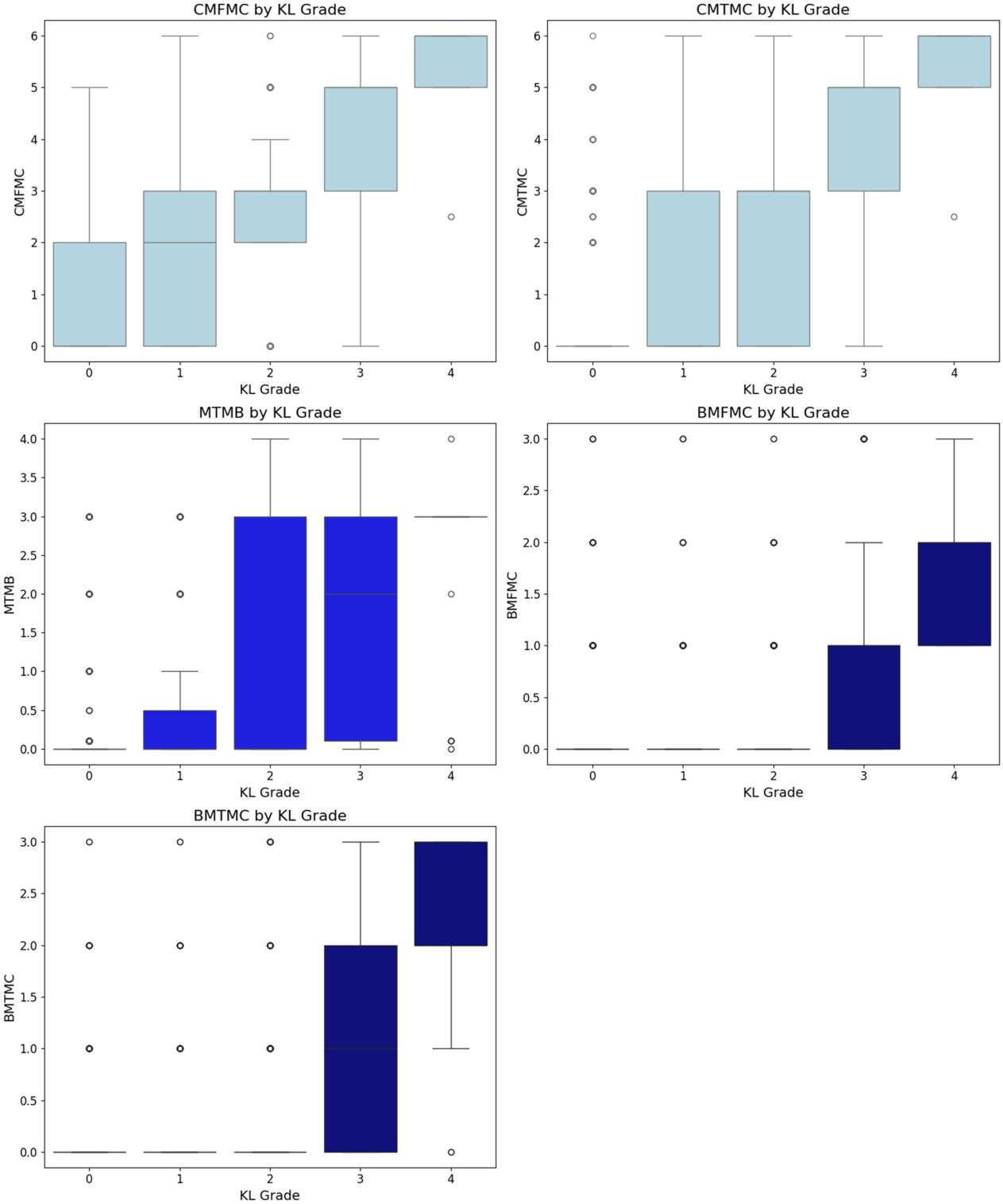
MRI findings of cartilage, meniscus and bone marrow in the central compartment are compared by X-ray KL grade.
Do Weon Lee, Ji-Sahn Kim, Hyuk-Soo Han, Du Hyun Ro. Longitudinal correlation between X-ray and MRI findings in medial compartment knee osteoarthritis: Insights into early cartilage loss and structural changes. Knee Surgery, Sports Traumatology, Arthroscopy. 2026;34(6):2047–2056. https://doi.org/10.1002/ksa.70016. © 2025 The Author(s).
Original Figure Legend
Figure 2
The research question
Participants and Methods
Main Findings
Implications for clinical and surgical practice
Limitations
Let's put it briefly.
Which clinical question does this address?
This supports the complementary information provided by meniscal and cartilage findings on MRI and standing X-rays in joint-preservation planning.
Keywords
- cartilage
- knee
- magnetic resonance imaging
- osteoarthritis
- radiography
- Osteoarthritis, Knee
- Magnetic Resonance Imaging
- Cartilage, Articular
Abstract · Original text
Do Weon Lee, Ji-Sahn Kim, Hyuk-Soo Han, Du Hyun Ro. Longitudinal correlation between X-ray and MRI findings in medial compartment knee osteoarthritis: Insights into early cartilage loss and structural changes. Knee surgery, sports traumatology, arthroscopy : official journal of the ESSKA. 2026;34(6):2047-2056. doi:10.1002/ksa.70016.
PURPOSE
Knee osteoarthritis (OA) is a common joint disorder assessed using radiographic (X-ray) and magnetic resonance imaging (MRI). While X-rays are accessible, MRI provides detailed insights into meniscus and cartilage. Few studies have evaluated the correlation between X-ray and MRI findings in knee OA longitudinally. This study addresses this gap by investigating their relationship over time.
METHODS
The Multicenter Osteoarthritis Study (MOST) dataset, a public, longitudinal cohort study focusing on knee OA in older adults, was used. The analysis included 3710 knees with medial compartment OA from different follow-ups over the course of 7 years. X-ray findings were compared with MRI findings, encompassing cartilage, meniscal and bone pathologies.
RESULTS
In the central compartment, knee OA progression began with cartilage loss, followed by meniscal and bone pathology, while in the posterior compartment, meniscal changes preceded cartilage and bone lesions. Cartilage loss in the central femur was the earliest event, even in Kellgren-Lawrence grade 0 knees, preceding X-ray changes. Tibial osteophytes developed first on X-ray, followed by joint space narrowing and femoral osteophytes. Longitudinal regression identified meniscal extrusion, cartilage loss and meniscal tear as significant predictors of OA progression (p < 0.001), with meniscal extrusion being the strongest.
CONCLUSION
Knee OA progression differs by compartment, with cartilage loss initiating changes centrally and meniscal pathology leading posteriorly. Tibial osteophytes appear early on X-ray. Meniscal extrusion is the strongest predictor of OA progression, highlighting the importance of MRI in identifying early changes and guiding personalised management.
LEVEL OF EVIDENCE
Level II.
© 2025 The Author(s). Knee Surgery, Sports Traumatology, Arthroscopy published by John Wiley & Sons Ltd on behalf of European Society of Sports Traumatology, Knee Surgery and Arthroscopy.
The original paper.
Longitudinal correlation between X-ray and MRI findings in medial compartment knee osteoarthritis: Insights into early cartilage loss and structural changes
Do Weon Lee, Ji-Sahn Kim, Hyuk-Soo Han, Du Hyun Ro
Knee Surgery, Sports Traumatology, Arthroscopy · 2026 · online 2025