
2026 · Knee Surgery, Sports Traumatology, Arthroscopy
X線上のOA変化とMRI上の軟骨・半月板・骨の変化には、時間的にどのような関係があるか?
中央部では軟骨損傷後に半月板・骨の変化が、後方部では半月板変化が先に認められました。中央大腿軟骨の減少はKL 0でも認められました。
原図Figure 1
 図を拡大する
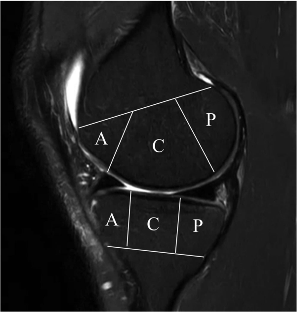
MRIでは軟骨と頭蓋骨を A の前に,C の真ん中に,P の後ろに分割します. 同じ膝は場所によって組織変化の量を変えることができますそこでレイアウトを分解しました
Do Weon Lee, Ji-Sahn Kim, Hyuk-Soo Han, Du Hyun Ro. Longitudinal correlation between X-ray and MRI findings in medial compartment knee osteoarthritis: Insights into early cartilage loss and structural changes. Knee Surgery, Sports Traumatology, Arthroscopy. 2026;34(6):2047–2056. https://doi.org/10.1002/ksa.70016. © 2025 The Author(s).
原著の図の説明
Figure 1
原図Figure 2
 図を拡大する
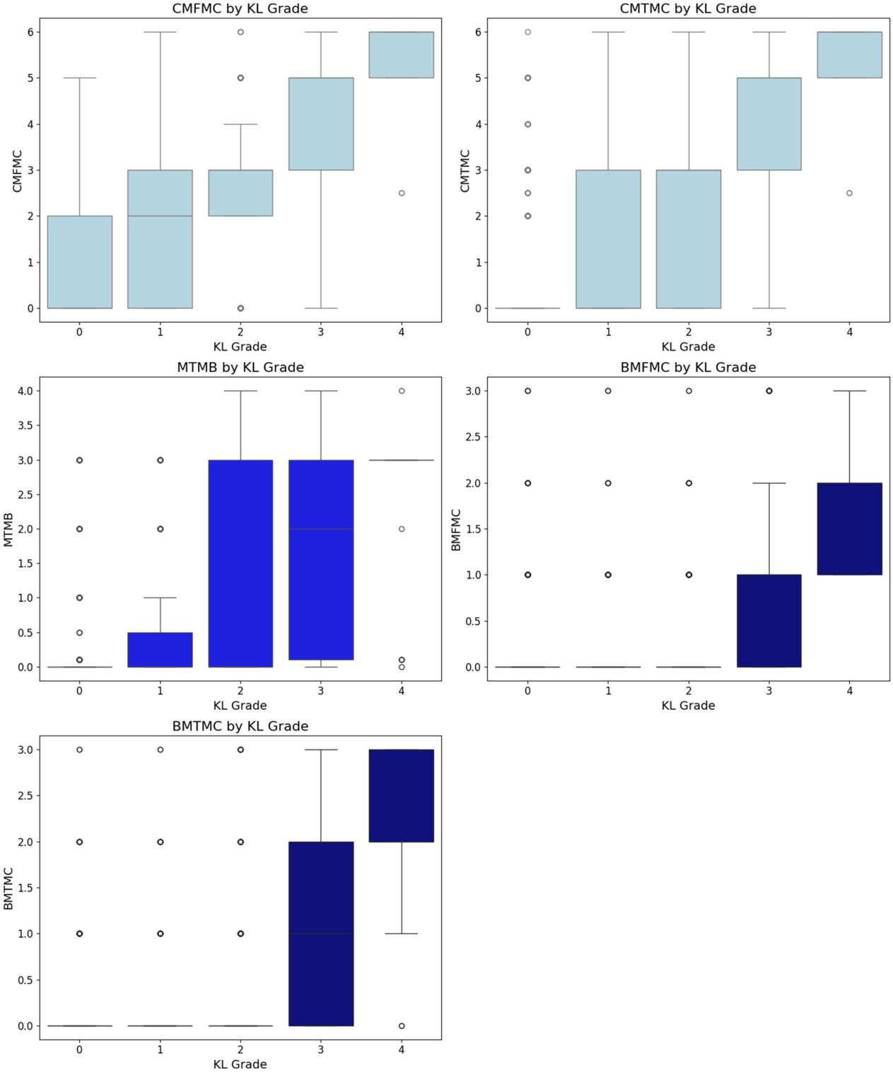
X線KLグレード別に、中央区画の軟骨・半月板・骨髄所見を比較しています。
Do Weon Lee, Ji-Sahn Kim, Hyuk-Soo Han, Du Hyun Ro. Longitudinal correlation between X-ray and MRI findings in medial compartment knee osteoarthritis: Insights into early cartilage loss and structural changes. Knee Surgery, Sports Traumatology, Arthroscopy. 2026;34(6):2047–2056. https://doi.org/10.1002/ksa.70016. © 2025 The Author(s).
原著の図の説明
Figure 2
研究で問うこと
対象と方法
主な結果
診療・手術で考える点
解釈の限界
簡単に説明しましょう
どの診療上の問いにつながりますか?
関節温存計画で、MRIの半月板・軟骨所見と立位X線が異なる情報を提供する根拠です。
Keywords・キーワード
- cartilage
- knee
- magnetic resonance imaging
- osteoarthritis
- radiography
- Osteoarthritis, Knee
- Magnetic Resonance Imaging
- Cartilage, Articular
Abstract・原文の抄録
Do Weon Lee, Ji-Sahn Kim, Hyuk-Soo Han, Du Hyun Ro. Longitudinal correlation between X-ray and MRI findings in medial compartment knee osteoarthritis: Insights into early cartilage loss and structural changes. Knee surgery, sports traumatology, arthroscopy : official journal of the ESSKA. 2026;34(6):2047-2056. doi:10.1002/ksa.70016.
PURPOSE
Knee osteoarthritis (OA) is a common joint disorder assessed using radiographic (X-ray) and magnetic resonance imaging (MRI). While X-rays are accessible, MRI provides detailed insights into meniscus and cartilage. Few studies have evaluated the correlation between X-ray and MRI findings in knee OA longitudinally. This study addresses this gap by investigating their relationship over time.
METHODS
The Multicenter Osteoarthritis Study (MOST) dataset, a public, longitudinal cohort study focusing on knee OA in older adults, was used. The analysis included 3710 knees with medial compartment OA from different follow-ups over the course of 7 years. X-ray findings were compared with MRI findings, encompassing cartilage, meniscal and bone pathologies.
RESULTS
In the central compartment, knee OA progression began with cartilage loss, followed by meniscal and bone pathology, while in the posterior compartment, meniscal changes preceded cartilage and bone lesions. Cartilage loss in the central femur was the earliest event, even in Kellgren-Lawrence grade 0 knees, preceding X-ray changes. Tibial osteophytes developed first on X-ray, followed by joint space narrowing and femoral osteophytes. Longitudinal regression identified meniscal extrusion, cartilage loss and meniscal tear as significant predictors of OA progression (p < 0.001), with meniscal extrusion being the strongest.
CONCLUSION
Knee OA progression differs by compartment, with cartilage loss initiating changes centrally and meniscal pathology leading posteriorly. Tibial osteophytes appear early on X-ray. Meniscal extrusion is the strongest predictor of OA progression, highlighting the importance of MRI in identifying early changes and guiding personalised management.
LEVEL OF EVIDENCE
Level II.
© 2025 The Author(s). Knee Surgery, Sports Traumatology, Arthroscopy published by John Wiley & Sons Ltd on behalf of European Society of Sports Traumatology, Knee Surgery and Arthroscopy.
オリジナル紙だ
Longitudinal correlation between X-ray and MRI findings in medial compartment knee osteoarthritis: Insights into early cartilage loss and structural changes
Do Weon Lee, Ji-Sahn Kim, Hyuk-Soo Han, Du Hyun Ro
Knee Surgery, Sports Traumatology, Arthroscopy · 2026 · online 2025