
2026 · Knee Surgery, Sports Traumatology, Arthroscopy
단순영상의 OA 변화와 MRI의 연골·반월연골·뼈 변화는 시간적으로 어떤 관계를 보일까?
중앙부에서는 연골손상 후 반월상연골·골 변화가, 후방부에서는 반월상연골 변화가 먼저 관찰됐습니다. 중앙 대퇴 연골 소실은 KL 0에서도 관찰됐습니다.
원문 Figure 1
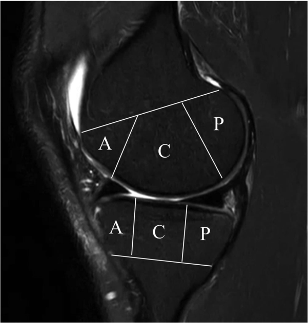 그림 크게 보기
MRI에서 대퇴골과 경골을 앞(A)·가운데(C)·뒤(P)로 나눕니다. 같은 무릎도 위치에 따라 조직 변화의 양상이 달라질 수 있어 구획을 나누어 분석했습니다.
Do Weon Lee, Ji-Sahn Kim, Hyuk-Soo Han, Du Hyun Ro. Longitudinal correlation between X-ray and MRI findings in medial compartment knee osteoarthritis: Insights into early cartilage loss and structural changes. Knee Surgery, Sports Traumatology, Arthroscopy. 2026;34(6):2047–2056. https://doi.org/10.1002/ksa.70016. © 2025 The Author(s).
원문 캡션
Figure 1
원문 Figure 2
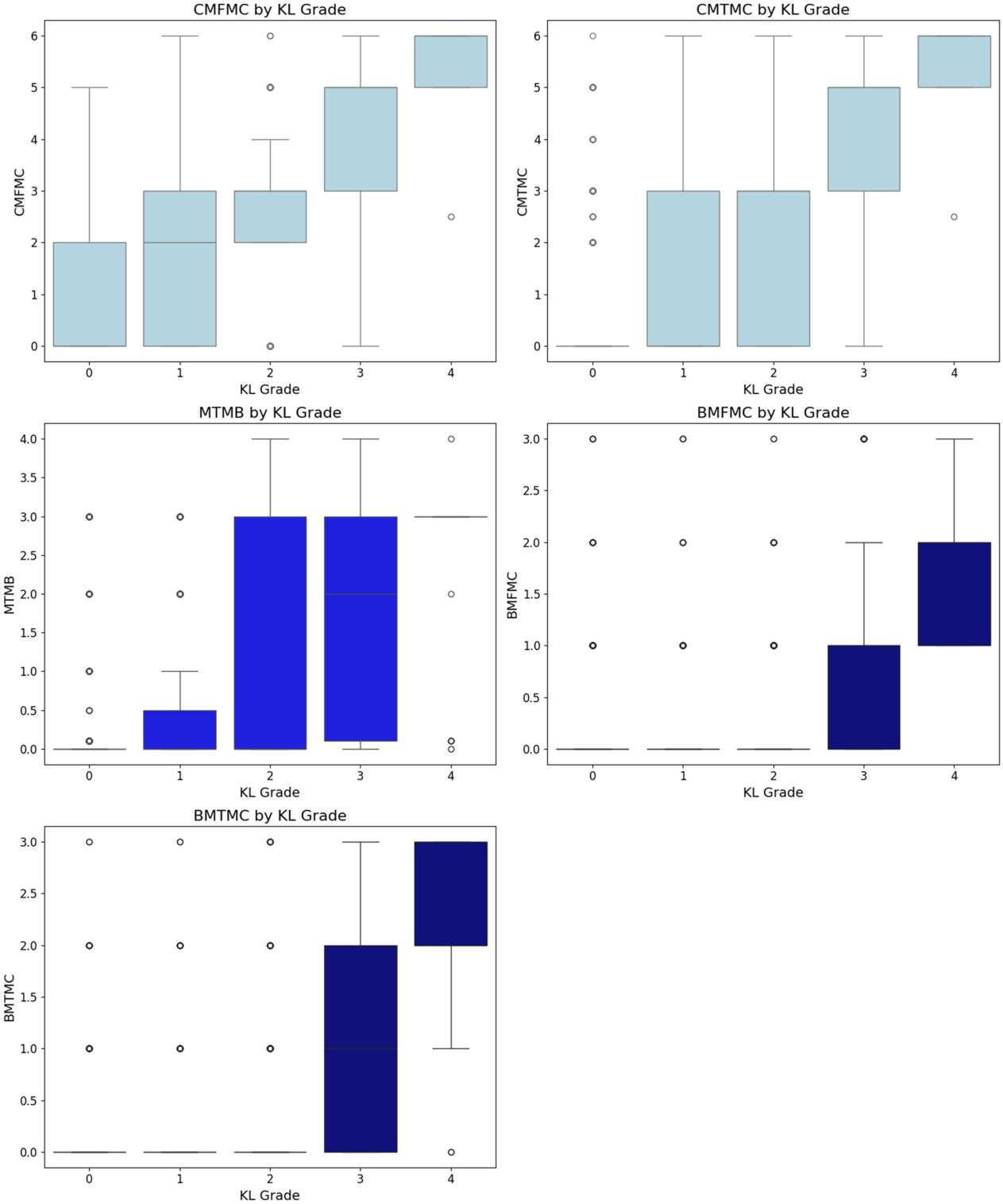 그림 크게 보기
X-ray KL 등급별 중앙 구획의 연골·연골판·골수 MRI 소견을 비교합니다.
Do Weon Lee, Ji-Sahn Kim, Hyuk-Soo Han, Du Hyun Ro. Longitudinal correlation between X-ray and MRI findings in medial compartment knee osteoarthritis: Insights into early cartilage loss and structural changes. Knee Surgery, Sports Traumatology, Arthroscopy. 2026;34(6):2047–2056. https://doi.org/10.1002/ksa.70016. © 2025 The Author(s).
원문 캡션
Figure 2
연구에서 묻는 질문
대상과 방법
주요 결과
진료와 수술에서 읽을 지점
해석의 제한
짧게 정리하면
어떤 진료 질문과 연결되나요?
관절 보존 계획에서 MRI의 반월연골·연골 소견과 기립 X-ray가 서로 다른 정보를 제공한다는 근거입니다.
Keywords · 주제어
- cartilage
- knee
- magnetic resonance imaging
- osteoarthritis
- radiography
- Osteoarthritis, Knee
- Magnetic Resonance Imaging
- Cartilage, Articular
Abstract · 원문 초록
Do Weon Lee, Ji-Sahn Kim, Hyuk-Soo Han, Du Hyun Ro. Longitudinal correlation between X-ray and MRI findings in medial compartment knee osteoarthritis: Insights into early cartilage loss and structural changes. Knee surgery, sports traumatology, arthroscopy : official journal of the ESSKA. 2026;34(6):2047-2056. doi:10.1002/ksa.70016.
PURPOSE
Knee osteoarthritis (OA) is a common joint disorder assessed using radiographic (X-ray) and magnetic resonance imaging (MRI). While X-rays are accessible, MRI provides detailed insights into meniscus and cartilage. Few studies have evaluated the correlation between X-ray and MRI findings in knee OA longitudinally. This study addresses this gap by investigating their relationship over time.
METHODS
The Multicenter Osteoarthritis Study (MOST) dataset, a public, longitudinal cohort study focusing on knee OA in older adults, was used. The analysis included 3710 knees with medial compartment OA from different follow-ups over the course of 7 years. X-ray findings were compared with MRI findings, encompassing cartilage, meniscal and bone pathologies.
RESULTS
In the central compartment, knee OA progression began with cartilage loss, followed by meniscal and bone pathology, while in the posterior compartment, meniscal changes preceded cartilage and bone lesions. Cartilage loss in the central femur was the earliest event, even in Kellgren-Lawrence grade 0 knees, preceding X-ray changes. Tibial osteophytes developed first on X-ray, followed by joint space narrowing and femoral osteophytes. Longitudinal regression identified meniscal extrusion, cartilage loss and meniscal tear as significant predictors of OA progression (p < 0.001), with meniscal extrusion being the strongest.
CONCLUSION
Knee OA progression differs by compartment, with cartilage loss initiating changes centrally and meniscal pathology leading posteriorly. Tibial osteophytes appear early on X-ray. Meniscal extrusion is the strongest predictor of OA progression, highlighting the importance of MRI in identifying early changes and guiding personalised management.
LEVEL OF EVIDENCE
Level II.
© 2025 The Author(s). Knee Surgery, Sports Traumatology, Arthroscopy published by John Wiley & Sons Ltd on behalf of European Society of Sports Traumatology, Knee Surgery and Arthroscopy.
원래 논문
Longitudinal correlation between X-ray and MRI findings in medial compartment knee osteoarthritis: Insights into early cartilage loss and structural changes
Do Weon Lee, Ji-Sahn Kim, Hyuk-Soo Han, Du Hyun Ro
Knee Surgery, Sports Traumatology, Arthroscopy · 2026 · online 2025