2026 · Knee Surgery & Related Research
Can Changing the Radiographic Angle Change the Measurement of the Same Knee?
The ICC between AI and expert measurements was 0.78 (95% CI 0.73–0.82). PTS increased by approximately 0.2° per 1° of internal rotation (R² = 0.43).
Original Figure 3
 Enlarge Figure
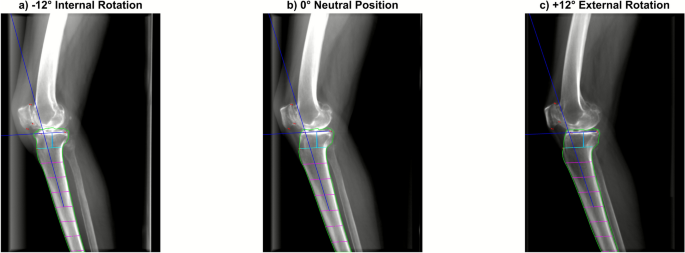
DRRs generated from one CT at different rotation angles, with automated PTS measurements.
Jaeseok Park, Andreas Persson, R Kyle Martin, Eivind Inderhaug, Sung Eun Kim, Sangyoon Kim, Donghyuk Kwak, Du Hyun Ro. Effects of axial malrotation on posterior tibial slope measurement: a digitally reconstructed radiograph study enabling automated quality assessment. Knee Surgery & Related Research (2026). https://doi.org/10.1186/s43019-026-00303-x
Participants and Methods
Main Findings
The point of reading on surgery
Limitations
Let's put it briefly.
What did this study confirm?
The ICC between AI and expert measurements was 0.78 (95% CI 0.73–0.82). PTS increased by approximately 0.2° per 1° of internal rotation (R² = 0.43).
Can it be applied directly to individual patients' treatment decisions?
This is a DRR-based proof of concept rather than a study of repeated clinical radiographs. Coronal tilt was fixed, and some measurement outliers occurred. Generalization to young ACL patients is limited.
Keywords
- Artificial intelligence
- Axial malrotation
- Digitally reconstructed radiograph
- Lateral knee radiograph
- Posterior tibial slope
- posterior tibial slope
- HTO
- ACL
Abstract · Original text
Jaeseok Park, Andreas Persson, R Kyle Martin, Eivind Inderhaug, Sung Eun Kim, Sangyoon Kim, Donghyuk Kwak, Du Hyun Ro. Effects of axial malrotation on posterior tibial slope measurement: a digitally reconstructed radiograph study enabling automated quality assessment. Knee surgery & related research. 2026;38(1):3. doi:10.1186/s43019-026-00303-x.
BACKGROUND
Accurate measurement of posterior tibial slope (PTS) is highly sensitive to axial malrotation of the knee during acquisition, but its impact has not been systematically quantified across different anatomical variations. This simulation study aimed to quantify the effect of axial malrotation on PTS using digitally reconstructed radiographs (DRRs) and suggest a practical marker for filtering out low-quality images with excessive malrotation.
MATERIALS AND METHODS
A total of 55 preoperative computed tomography (CT) scans from January 2021 to December 2024 in a single, tertiary hospital were retrospectively reviewed. DRRs were generated from those scans to simulate lateral knee radiographs with malrotation ranging from -12° to +12° relative to an anatomically aligned baseline. An artificial-intelligence (AI)-based tool automatically measured PTS on each DRR, with agreement evaluated using intraclass correlation coefficient (ICC). PCDR was calculated from femoral contours and analyzed for correlation with malrotation angles and resulting PTS measurement error.
RESULTS
AI-based PTS measurements on DRRs showed good agreement with expert annotations (ICC = 0.78, 95% CI 0.73-0.82). PTS increased linearly with internal rotation, with each 1° of rotation resulting in approximately 0.2° change in PTS (R2 = 0.43, p < 0.01). Errors exceeded 1° when malrotation surpassed ±6°. PCDR was strongly correlated with malrotation angle (R2 > 0.98, p < 0.001) and achieved fair discriminative performance as a binary classifier for > 1° PTS error [area under the receiver operating curve (AUROC) = 0.77].
CONCLUSIONS
CT-derived DRRs combined with AI analysis showed that PTS measurement error proportionately increased with axial malrotation. Identifying and excluding radiographs with excessive rotation improves the reliability of slope-based assessments and supports more accurate surgical planning.
LEVEL OF EVIDENCE
III, retrospective cohort study.
© The Author(s) 2026
The original paper.
Effects of axial malrotation on posterior tibial slope measurement: a digitally reconstructed radiograph study enabling automated quality assessment
Jaeseok Park, Andreas Persson, R Kyle Martin, Eivind Inderhaug, Sung Eun Kim, Sangyoon Kim, Donghyuk Kwak, Du Hyun Ro
Knee Surgery & Related Research · 2026