2025 · Knee Surgery & Related Research
How Are Muscle-activation Changes in PFPS Related to the Sagittal Knee Moment during Walking?
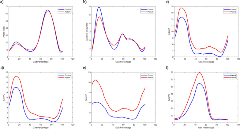
The PFPS group had lower knee-extension moment during weight acceptance, without a significant difference in flexion angle. ST activity was higher throughout gait.
Fig. 1 is the original.
 Enlarge Figure
Blue lines show control-group means and red lines show PFPS-group means across the gait cycle. Angles, knee-extension moments, and EMG from four muscles are aligned on the same time axis.
Byung Sun Choi, Soon Bin Kwon, Sehyeon Jeon, Myeongjun Kim, Yunseo Ku, Du Hyun Ro, Hyuk-Soo Han. Relationship between muscle activation and sagittal knee joint biomechanics in patients with patellofemoral pain syndrome: a cross-sectional study. Knee Surgery & Related Research (2025). DOI: 10.1186/s43019-025-00259-4. © The Author(s) 2025
Original Figure Legend
Kinetic, kinematic, and EMG data. All curves represent the mean values of biomechanical features at the point of the gait …
Participants and Methods
The order in which you read the painting.
Main Findings
The point of reading on surgery
Limitations
Let's put it briefly.
What did this study confirm?
The PFPS group had lower knee-extension moment during weight acceptance, without a significant difference in flexion angle. ST activity was higher throughout gait.
Can it be applied directly to individual patients' treatment decisions?
This is a cross-sectional comparison. The compensatory mechanism is an interpretation, not proof of causality. Rehabilitation and surgical interventions were not compared.
Keywords
- Electromyography
- Gait analysis
- Patellofemoral pain syndrome
- PFJ
- patellofemoral pain
- gait
- EMG
Abstract · Original text
Byung Sun Choi, Soon Bin Kwon, Sehyeon Jeon, Myeongjun Kim, Yunseo Ku, Du Hyun Ro, Hyuk-Soo Han. Relationship between muscle activation and sagittal knee joint biomechanics in patients with patellofemoral pain syndrome: a cross-sectional study. Knee surgery & related research. 2025;37(1):7. doi:10.1186/s43019-025-00259-4.
BACKGROUND
Patellofemoral pain syndrome (PFPS) is one of the most common conditions affecting the knee joint, yet its pathomechanics remain unclear. The aim of this study was to investigate changes in muscle activation and gait patterns and to analyze the relationship between muscle activation and kinetic gait patterns in patients with PFPS.
METHODS
This study included 31 patients with PFPS and 28 healthy volunteers without any symptoms. The sagittal plane motion of the knee joint, representing primary movement of the knee joint, was evaluated to identify changes in gait patterns. Electromyography (EMG) was used to measure muscle activation of vastus medialis (VM), vastus lateralis (VL), semitendinosus (ST), and gastrocnemius (GCM) muscles during gait analysis. Biomechanical features were analyzed during the three phases of the gait cycle; weight acceptance (WA), single limb support (SLS), and swing limb advancement (SLA) (0 ~ 12%, 13 ~ 50%, and 51 ~ 100% of the gait cycle, respectively).
RESULTS
The average knee extension moment (KEM) during WA was lower in the patient group and no significant differences were observed in the knee flexion angle (KFA). With respect to muscle activation, the patient group showed significantly higher muscle activation of the ST muscle in all phases. As the absolute value of the moment increased, the activation of the VM, VL, and ST muscles increased more rapidly in the patient group, especially when KEM was under -1% body weight × height (Bw × Ht) or over 5% Bw × Ht.
CONCLUSIONS
Patients with PFPS exhibit elevated muscle activation, particularly in response to changes in the knee extension moment, which is likely a compensatory mechanism to manage knee joint loading during gait. These results highlight altered neuromuscular adaptations in PFPS, suggesting targeted therapies may help improve functional outcomes. Level of evidence III, cross-sectional study.
© The Author(s) 2025
The original paper.
Relationship between muscle activation and sagittal knee joint biomechanics in patients with patellofemoral pain syndrome: a cross-sectional study
Byung Sun Choi, Soon Bin Kwon, Sehyeon Jeon, Myeongjun Kim, Yunseo Ku, Du Hyun Ro, Hyuk-Soo Han
Knee Surgery & Related Research · 2025