2024 · Skeletal Radiology
Does a ±5° Beam-angle Difference Change Skyline PFJ Measurements, and Can the Anterior Tibial Border Guide Imaging?
Sulcus angle, congruence angle, patellar tilt, lateral facet angle and bisect ratio did not differ significantly across beam angles (all P > 0.05).
Fig. 3 is the original.
 Enlarge Figure
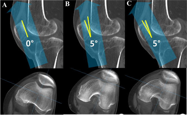
Skyline images reconstructed from the same CT with the beam at neutral, 5° upward and 5° downward.
Sung Eun Kim, Sunghyun Hwang, Ji Han Lee, Geunwu Gimm, Hyuk-Soo Han, Byung Sun Choi, Du Hyun Ro. Minimal impact of beam projection angle deviations in skyline (Laurin) view and the efficacy of the anterior border of proximal tibia as a guiding landmark. Skeletal Radiology (2024). DOI: 10.1007/s00256-024-04619-1. © The Author(s) 2024
Original Figure Legend
Examples of beam projection angle deviations and corresponding digitally reconstructed skyline images from CT scans. A Neutral tilt, B 5° …
Participants and Methods
The order in which you read the painting.
Main Findings
The point of reading on surgery
Limitations
Let's put it briefly.
What did this study confirm?
Sulcus angle, congruence angle, patellar tilt, lateral facet angle and bisect ratio did not differ significantly across beam angles (all P > 0.05).
Can it be applied directly to individual patients' treatment decisions?
The findings are limited to the tested acquisition conditions and the ±5° range. Larger angle errors, severe deformity, and improvements in clinical diagnosis or surgical outcomes were not directly assessed.
Keywords
- Anterior border of proximal tibia
- Beam projection angle
- Deviation
- Laurin view
- Skyline view
- Tibia
- Tomography, X-Ray Computed
- Retrospective Studies
Abstract · Original text
Sung Eun Kim, Sunghyun Hwang, Ji Han Lee, Geunwu Gimm, Hyuk-Soo Han, Byung Sun Choi, Du Hyun Ro. Minimal impact of beam projection angle deviations in skyline (Laurin) view and the efficacy of the anterior border of proximal tibia as a guiding landmark. Skeletal radiology. 2024;53(7):1399-1406. doi:10.1007/s00256-024-04619-1.
OBJECTIVE
Obtaining an optimal knee skyline view is challenging due to inaccuracies in beam projection angles (BPAs) and soft tissue obscuring bony landmarks. This study aimed to assess the impact of BPA deviations on patellofemoral index measurements and assessed the anterior border of the proximal tibia as an anatomic landmark for guiding BPAs.
MATERIALS AND METHODS
This retrospective study consisted of three parts. The first was a simulation study using 52 CT scans of knees with a 20° flexion contracture to replicate the skyline (Laurin) view. Digitally reconstructed radiographs simulated neutral, 5° downward, and 5° upward tilt BPAs. Five patellofemoral indices (sulcus angle, congruence angle, patellar tilt angle, lateral facet angle, and bisect ratio) were measured and compared. The second part was a proof of concept study on 162 knees to examine patellar indices differences across these BPAs. Lastly, the alignment of the anterior border of the proximal tibia with the BPA tangential to the patellar articular surface was tested from the CT scans.
RESULTS
No significant differences in patellofemoral indices were found across various BPAs in both the simulation and proof of concept studies (all p > 0.05). The angle between the anterior border of the proximal tibia and the patellar articular surface was 1.5 ± 5.3°, a statistically significant (p = 0.037) yet clinically acceptable deviation.
CONCLUSION
Patellofemoral indices in skyline view remained consistent regardless of BPA deviations. The anterior border of the proximal tibia proved to be an effective landmark for accurate beam projection.
© The Author(s) 2024
The original paper.
Minimal impact of beam projection angle deviations in skyline (Laurin) view and the efficacy of the anterior border of proximal tibia as a guiding landmark
Sung Eun Kim, Sunghyun Hwang, Ji Han Lee, Geunwu Gimm, Hyuk-Soo Han, Byung Sun Choi, Du Hyun Ro
Skeletal Radiology · 2024