2024 · Skeletal Radiology
スカイラインX線のビーム角度±5°の差でPFJ指標は変わるか、また脛骨前縁は撮影基準となるか?
sulcus angle・congruence angle・patellar tilt・lateral facet angle・bisect ratioは、ビーム角度間で有意差がありませんでした(すべてP > 0.05)。
図3はオリジナルです
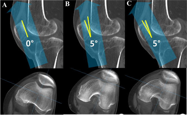 図を拡大する
同一CTから、ビームを中立・上方5°・下方5°に調整して再構成したskyline画像です。
Sung Eun Kim, Sunghyun Hwang, Ji Han Lee, Geunwu Gimm, Hyuk-Soo Han, Byung Sun Choi, Du Hyun Ro. Minimal impact of beam projection angle deviations in skyline (Laurin) view and the efficacy of the anterior border of proximal tibia as a guiding landmark. Skeletal Radiology (2024). DOI: 10.1007/s00256-024-04619-1. © The Author(s) 2024
原著の図の説明
Examples of beam projection angle deviations and corresponding digitally reconstructed skyline images from CT scans. A Neutral tilt, B 5° …
対象と方法
絵を読んだ順序です
主な結果
手術に関する読書のポイント
解釈の限界
簡単に説明しましょう
この研究で何が確認されましたか?
sulcus angle・congruence angle・patellar tilt・lateral facet angle・bisect ratioは、ビーム角度間で有意差がありませんでした(すべてP > 0.05)。
治療決定に直接適用できるか?
検証した撮影条件と±5°の範囲に限られます。より大きな角度誤差、重度変形の影響、臨床診断や手術結果の改善は直接評価していません。
Keywords・キーワード
- Anterior border of proximal tibia
- Beam projection angle
- Deviation
- Laurin view
- Skyline view
- Tibia
- Tomography, X-Ray Computed
- Retrospective Studies
Abstract・原文の抄録
Sung Eun Kim, Sunghyun Hwang, Ji Han Lee, Geunwu Gimm, Hyuk-Soo Han, Byung Sun Choi, Du Hyun Ro. Minimal impact of beam projection angle deviations in skyline (Laurin) view and the efficacy of the anterior border of proximal tibia as a guiding landmark. Skeletal radiology. 2024;53(7):1399-1406. doi:10.1007/s00256-024-04619-1.
OBJECTIVE
Obtaining an optimal knee skyline view is challenging due to inaccuracies in beam projection angles (BPAs) and soft tissue obscuring bony landmarks. This study aimed to assess the impact of BPA deviations on patellofemoral index measurements and assessed the anterior border of the proximal tibia as an anatomic landmark for guiding BPAs.
MATERIALS AND METHODS
This retrospective study consisted of three parts. The first was a simulation study using 52 CT scans of knees with a 20° flexion contracture to replicate the skyline (Laurin) view. Digitally reconstructed radiographs simulated neutral, 5° downward, and 5° upward tilt BPAs. Five patellofemoral indices (sulcus angle, congruence angle, patellar tilt angle, lateral facet angle, and bisect ratio) were measured and compared. The second part was a proof of concept study on 162 knees to examine patellar indices differences across these BPAs. Lastly, the alignment of the anterior border of the proximal tibia with the BPA tangential to the patellar articular surface was tested from the CT scans.
RESULTS
No significant differences in patellofemoral indices were found across various BPAs in both the simulation and proof of concept studies (all p > 0.05). The angle between the anterior border of the proximal tibia and the patellar articular surface was 1.5 ± 5.3°, a statistically significant (p = 0.037) yet clinically acceptable deviation.
CONCLUSION
Patellofemoral indices in skyline view remained consistent regardless of BPA deviations. The anterior border of the proximal tibia proved to be an effective landmark for accurate beam projection.
© The Author(s) 2024
オリジナル紙だ
Minimal impact of beam projection angle deviations in skyline (Laurin) view and the efficacy of the anterior border of proximal tibia as a guiding landmark
Sung Eun Kim, Sunghyun Hwang, Ji Han Lee, Geunwu Gimm, Hyuk-Soo Han, Byung Sun Choi, Du Hyun Ro
Skeletal Radiology · 2024