What Limits the Clinical Adoption of Orthopedic AI, and What Validation Is Needed?
This narrative review compares perioperative risk prediction, fracture and OA imaging, and LLM support for documentation and communication. It examines the validation and operational steps needed to turn model accuracy into clinical benefit.
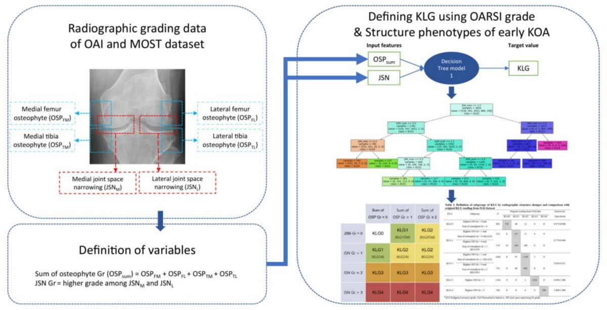
A diagram linking osteophytes and joint-space narrowing to KL grades.
Sang Yoon Kim, Byung Sun Choi, Hyuk-Soo Han, Du Hyun Ro. Artificial intelligence in orthopedics: current applications, challenges, and future directions. Knee Surgery & Related Research (2026). DOI: 10.1186/s43019-026-00317-5 · Figure 3. Figure created by the article authors based on Ko et al.
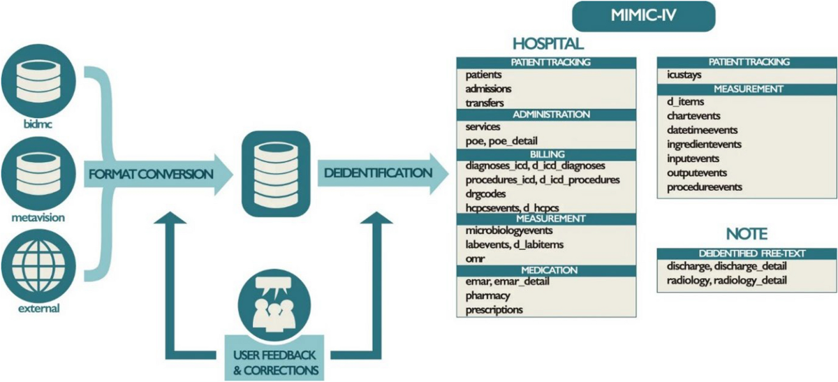
MIMIC-IV links hospital, intensive care and clinical-document data.
Sang Yoon Kim, Byung Sun Choi, Hyuk-Soo Han, Du Hyun Ro. Artificial intelligence in orthopedics: current applications, challenges, and future directions. Knee Surgery & Related Research (2026). DOI: 10.1186/s43019-026-00317-5 · Figure 1. Adapted from Johnson et al. under CC BY 4.0; changes were made by the article authors.
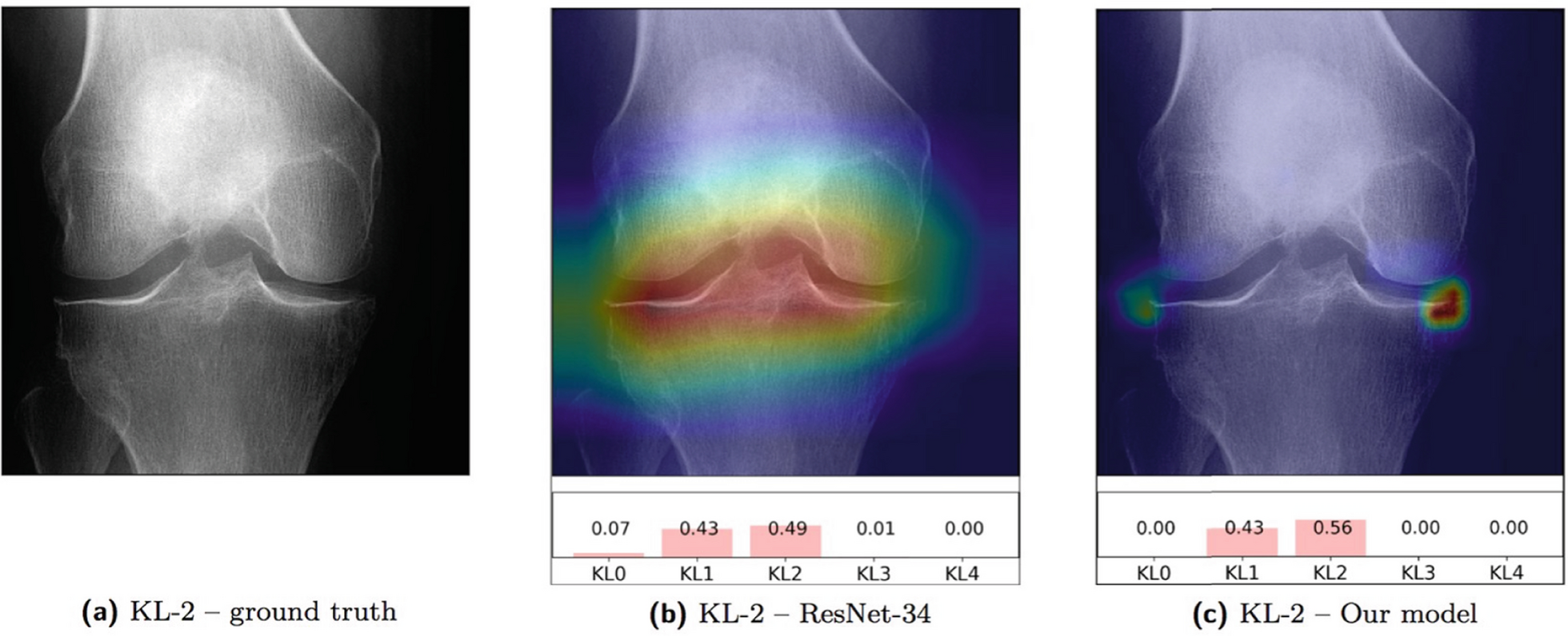
Chain-of-thought (CoT) rationale for automated Kellgren–Lawrence (KL) grading. a Input anteroposterior weight-bearing radiograph; b Standard Grad-CAM saliency showing nonspecific feature …
The review question and evidence scope
High retrospective accuracy does not necessarily improve routine care. The review separates structured-data machine learning, imaging deep learning and text-based LLMs to identify tasks approaching deployment and unresolved barriers.
It purposively selected mainly 2020–2025 studies emphasizing external validation, prospective assessment, workflow integration or multimodal and implementation methods. It did not test a new cohort or model and is not an exhaustive systematic review or pooled-effect meta-analysis.
Matching data and models to the clinical task
Structured EMR variables such as age, hemoglobin, comorbidities and medication suit logistic regression, random forests and boosting. Pixel-based classification, segmentation and measurement suit deep learning; operative notes, reports and discharge drafts suit language models.
Complexity alone is not a reason to choose a model. Define available inputs, prediction time and the clinical endpoint, compare with simpler baselines, and establish the added value of multimodal fusion over individual inputs.
Perioperative prediction must inform a clinical action
The review discusses transfusion, early complications and 30-day readmission after arthroplasty. The cited transfusion study reported internal and external AUROC of 0.842 and 0.880; these are results from that study, not a new analysis by the review.
Use requires calibrated probabilities and actionable thresholds. For transfusion risk, assess how predictions affect anemia evaluation, blood preparation or monitoring and the balance of unnecessary actions and missed cases. Discrimination alone does not establish useful clinical action.
Imaging AI for diagnostic support and standardized measurement
Fracture detection, KL grading, alignment and posterior tibial slope measurement can standardize repetitive work. Distinguish direct composite grading from feature-based pipelines that measure osteophytes and joint-space narrowing before mapping to a grade; original Figure 3 illustrates this logic.
Metrics must match the task: sensitivity and missed lesions for diagnosis, error and agreement for angles, and overlap and boundary accuracy for segmentation. Robustness across equipment, positioning and severity, and improvement with clinician assistance require separate evaluation.
LLMs for documentation, extraction and communication
LLMs can summarize histories, extract structured information from operative or imaging reports, and draft clinician-reviewed explanations and discharge instructions. Examination performance does not establish reliable diagnosis or treatment in real patients.
Evaluate hallucination, omission, prompt sensitivity, malicious document instructions and privacy risks. Retrieval grounding, constrained outputs, intended-use boundaries and clinician review matter; assess editing burden, missing critical information, comprehension and safety alongside documentation time.
Leakage prevention and external validation
Separate development and test data at least by patient. Mixing bilateral knees or repeat images across sets can inflate apparent generalization, and inputs must be available at the intended prediction time.
External validation tests different institutions, periods, equipment and patient subgroups rather than merely increasing sample size. Prespecify and disclose performance and calibration failures and unsupported cases instead of relying on a single average metric.
Discrimination, calibration and net benefit answer different questions
Discrimination separates higher- from lower-risk patients; calibration evaluates whether predicted probabilities match observed frequencies. AUROC alone cannot show whether a predicted 20% risk corresponds to approximately 20% events. Assess calibration plots and measures such as the Brier score.
Decision curves compare net benefit across actionable thresholds, balancing unnecessary action and missed cases. Retrospective net benefit does not prove improved outcomes; prospective evaluation must examine decisions and safety.
How to interpret explainability figures
Saliency and Grad-CAM visualize image regions associated with a model output. Original Figure 5 shows an X-ray, saliency map and KL grade probabilities together.
From silent-mode evaluation to clinician–AI assessment
Silent-mode deployment can reveal local input errors, missingness and performance degradation without changing clinical decisions. Subsequent clinician-facing assessment should measure time, editing, automation bias and handling of exceptions.
Standalone accuracy differs from clinician–AI performance. Comparative trials, staged rollout and pragmatic prospective studies should assess workload and patient-centered outcomes. The review highlights transparent frameworks including DECIDE-AI and CONSORT-AI.
Monitoring and accountability after deployment
Changes in testing, case mix or practice can erode performance. Record versions and changes; monitor input distributions, calibration, subgroup performance and failures; define escalation and stop-use conditions.
EMR/PACS integration and interface design are operational requirements beyond accuracy. Minimize duplicate input and alert burden while exposing limitations, delineate responsibility across developers, vendors, institutions and users, and audit unequal subgroup effects.
Multimodal models and digital twins: promise and evidence gaps
The review proposes future multimodal representations linking imaging phenotypes, clinical variables and intraoperative data to patient trajectories, potentially providing complementary information for progression or revision prediction.
It does not validate an operational digital twin or its benefit. Reliable linkage, missingness, cross-site variation, leakage prevention, incremental value and prospective net benefit remain requirements; these concepts do not establish a patient's future with certainty.
Applying the review in practice
For an adoption decision, examine intended use and patients, inputs and timing, external validation and calibration, clinician-assisted benefit, exception handling and ongoing monitoring. Evaluate measurement reliability, efficiency, understanding, decisions and patient outcomes as distinct endpoints.
The review provides an adoption framework, not a guarantee for all products or a specific model. Individual decisions require the primary studies, current authorized intended use and local evaluation.
Let's put it briefly.
Which orthopedic AI tasks are nearer clinical use?
The review identifies structured risk prediction and imaging measurement as relatively nearer deployment, subject to product-specific external validation, workflow fit and demonstrated benefit with clinician use.
Does a high AUROC justify immediate use?
No. Evaluate calibration, thresholds, external and subgroup performance, missed cases and unnecessary actions, and test the tool prospectively in care.
Does an explainability image establish trustworthiness?
It offers clues, not proof of correct causal reasoning or safety. Examine actual failures, measured features and independent performance as well.
What is a practical near-term role for LLMs?
Clinician-reviewed summarization, extraction and drafting are practical roles. Assess missing critical facts, editing burden, privacy and incorrect advice alongside prose quality.
Do digital twins already predict patient trajectories with certainty?
No. This is a research direction in the review; linked-data reliability, incremental predictive value and prospective benefit require validation.
Keywords
Artificial intelligence
Clinical translation
Deep learning
External validation
Large language models
Machine learning
Musculoskeletal imaging
Orthopedics
Abstract · Original text
Sang Yoon Kim, Byung Sun Choi, Hyuk-Soo Han, Du Hyun Ro. Artificial intelligence in orthopedics: current applications, challenges, and future directions. Knee surgery & related research. 2026;38(1):14. doi:10.1186/s43019-026-00317-5.
BACKGROUND
Artificial intelligence research in orthopedics has grown rapidly, yet a substantial gap remains between technical development and clinical translation. This narrative review summarizes current applications of artificial intelligence in orthopedic practice and highlights barriers to implementation.
MAIN BODY
Current work converges on three domains: machine learning for structured perioperative risk prediction, deep learning for standardized musculoskeletal imaging, and large language models for workflow and decision support. Applications such as automated fracture detection, Kellgren-Lawrence grading for osteoarthritis, and transfusion risk modeling are approaching clinical maturity. However, routine adoption is limited by algorithmic opacity, performance degradation in new clinical environments, and poor fit within existing workflows. We argue that progress should shift from increasing model complexity toward rigorous evaluation, including external validation on independent cohorts. In addition, probability calibration and uncertainty estimation are important for trustworthy risk communication. Future directions may include multimodal "digital twin" approaches that integrate electronic medical records, imaging phenotypes, and intraoperative data into patient-specific trajectories.
CONCLUSIONS
The clinical impact of artificial intelligence in orthopedics will depend on life-cycle governance and demonstrated net benefit, prioritizing reliability and implementation science over retrospective benchmark performance.
 Enlarge Figure
 Enlarge Figure
 Enlarge Figure