2023 · KSSTA
전장 하지 X-ray의 15개 기준점을 찾으면 정렬 각도를 자동으로 재현할 수 있을까?
자동 모델의 각도별 inter-observer ICC는 ≥0.98, 반복 측정 ICC는 1.00이었습니다.
연구 수치로 읽는 결과
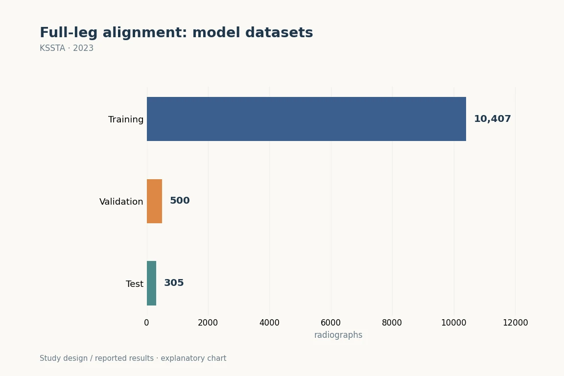 그림 크게 보기
학습 10,407장, 검증 500장, 시험 305장으로 구성한 단일 기관 연구입니다. 이 그림은 데이터 분리 구조를 보여줍니다. 세 기관 외부 검증은 후속 2024년 연구에서 따로 시행했습니다.
KSSTA (2023). DOI: 10.1007/s00167-022-07124-x · Original explanatory chart based on reported data
연구에서 묻는 질문
대상과 방법
주요 결과
진료와 수술에서 읽을 지점
해석의 제한
짧게 정리하면
어떤 진료 질문과 연결되나요?
HTO·TKA 계획의 출발점인 mLDFA/MPTA/HKAA 측정을 표준화하는 기반입니다. 자동 측정은 촬영 표준화, 관절 내 변형 해석 및 수술자의 최종 계획을 대신하지 않습니다.
Keywords · 주제어
- Angle measurement
- Deep learning
- Hip knee ankle angle (HKAA)
- Joint line convergence angle (JLCA)
- Landmark recognition
- Lower extremity
- Mechanical lateral distal femoral angle (mLDFA)
- Medial proximal tibial angle (MPTA)
Abstract · 원문 초록
Changwung Jo, Doohyun Hwang, Sunho Ko, Myung Ho Yang, Myung Chul Lee, Hyuk-Soo Han, Du Hyun Ro. Deep learning-based landmark recognition and angle measurement of full-leg plain radiographs can be adopted to assess lower extremity alignment. Knee surgery, sports traumatology, arthroscopy : official journal of the ESSKA. 2023;31(4):1388-1397. doi:10.1007/s00167-022-07124-x.
PURPOSE
Evaluating lower extremity alignment using full-leg plain radiographs is an essential step in diagnosis and treatment of patients with knee osteoarthritis. The study objective was to present a deep learning-based anatomical landmark recognition and angle measurement model, using full-leg radiographs, and validate its performance.
METHODS
A total of 11,212 full-leg plain radiographs were used to create the model. To train the data, 15 anatomical landmarks were marked by two orthopaedic surgeons. Mechanical lateral distal femoral angle (mLDFA), medial proximal tibial angle (MPTA), joint line convergence angle (JLCA), and hip-knee-ankle angle (HKAA) were then measured. For inter-observer reliability, the inter-observer intraclass correlation coefficient (ICC) was evaluated by comparing measurements from the model, surgeons, and students, to ground truth measurements annotated by an orthopaedic specialist with 14 years of experience. To evaluate test-retest reliability, all measurements were made twice by each measurer. Intra-observer ICCs were then derived. Performance evaluation metrics used in previous studies were also derived for direct comparison of the model's performance.
RESULTS
Inter-observer ICCs for all angles of the model were 0.98 or higher (p < 0.001). Intra-observer ICCs for all angles were 1.00, which was higher than that of the orthopaedic specialist (0.97-1.00). Measurements made by the model showed no significant systemic variation. Except for JLCA, angles were precisely measured with absolute error averages under 0.52 degrees and proportion of outliers under 4.26%.
CONCLUSIONS
The deep learning model is capable of evaluating lower extremity alignment with performance as accurate as an orthopaedic specialist with 14 years of experience.
LEVEL OF EVIDENCE
III, retrospective cohort study.
원래 논문
Deep learning-based landmark recognition and angle measurement of full-leg plain radiographs can be adopted to assess lower extremity alignment
Jo C, Hwang D, Ko S, Yang MH, Lee MC, Han HS, Ro DH
KSSTA · 2023 · online 2022