2026 · Bone & Joint Open
As OA Severity Increases, Do the Actual and Arithmetically Estimated Native Joint Lines Change in the Same Way?
In medial OA, TJLA changed from medial-downward to medial-upward at KL 3. TJLA changes were observed at KL 3–4 in both medial and lateral OA (P < 0.001).
Research question and design
 Enlarge Figure
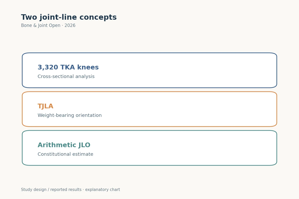
Weight-bearing joint-line angle (TJLA) was compared with arithmetic aJLO. TJLA differed in KL 3–4, while aJLO remained relatively stable.
Bone & Joint Open (2026). DOI: 10.1302/2633-1462.72.BJO-2025-0370.R1 · Original explanatory chart based on reported data
The research question
Participants and Methods
Main Findings
Implications for clinical and surgical practice
Limitations
Let's put it briefly.
Which clinical question does this address?
The findings support distinguishing weight-bearing TJLA from arithmetic aJLO in studies of individualized TKA alignment.
Keywords
- Tibiofemoral joint line
- Osteoarthritis severity
- Constitutional alignment
- Radiological analysis
- TKA
- alignment
- CPAK
- aJLO
Abstract · Original text
John E Farey, Benjamin J Leong, Jil A Wood, Do W Lee, Seyun Kim, Darren B Chen, Du H Ro, Samuel J MacDessi. Dynamic changes to the tibiofemoral joint line with increasing osteoarthritis severity and its relationship to constitutional alignment : a radiological analysis of 3,320 knees. Bone & joint open. 2026;7(2):249-258. doi:10.1302/2633-1462.72.BJO-2025-0370.R1.
AIMS
Changes in both native and dynamic joint line obliquity may influence the pathogenesis of osteoarthritis (OA) and subsequent development of coronal limb alignment deformity. This study aimed to characterize joint line alterations relative to OA severity and constitutional alignment.
METHODS
A retrospective, cross-sectional analysis of 3,320 preoperative weightbearing long-leg radiographs from patients undergoing total knee arthroplasty was performed using deep learning software. OA severity was assessed using the Kellgren-Lawrence (KL) grading system. Constitutional alignment parameters included lateral distal femoral angle (LDFA), medial proximal tibial angle (MPTA), arithmetic hip-knee-ankle (aHKA) angle, and arithmetic joint line obliquity (aJLO). Tibiofemoral joint line alterations were assessed using the mechanical hip-knee-ankle (mHKA) angle, tibial joint line angle (TJLA), femoral joint line angle (FJLA), and joint line congruence angle (JLCA). The primary outcome was the difference in the TJLA relative to the aJLO across KL grades, stratified for the presence of medial or lateral compartment OA.
RESULTS
Alterations in TJLA were observed in moderate (KL Grade 3) and severe (Grade 4) OA for medial and lateral compartment OA (both p < 0.001). In medial OA, the TJLA reversed orientation at Grade 3 from medially declined to medially inclined relative to aJLO. The aJLO maintained its constitutional orientation until Grade 4, when non-clinically significant changes (< 1°) were observed (p < 0.001). In lateral OA, the TJLA became more medially inclined in moderate and severe OA, while the aJLO remained stable. Significant changes in aHKA were observed in moderate and severe OA, indicating an apparent change in constitutional limb alignment (p < 0.001 for both medial and lateral OA).
CONCLUSION
The tibiofemoral joint line angle changes dynamically with OA progression, while the constitutional aJLO remains stable until advanced disease occurs. Understanding this distinction between constitutional and dynamic joint line orientation is important when communicating and studying outcomes related to knee alignment.
© 2026 Farey et al.
The original paper.
Dynamic changes to the tibiofemoral joint line with increasing osteoarthritis severity and its relationship to constitutional alignment: a radiological analysis of 3,320 knees
John E Farey, Benjamin J Leong, Jil A Wood, Do W Lee, Seyun Kim, Darren B Chen, Du Hyun Ro, Samuel J MacDessi
Bone & Joint Open · 2026