TKA · RESIDENTS & FELLOWS
Total Knee Arthroplasty
This section covers alignment planning, bone resection, soft-tissue balance, and fixation. It explains how each stage of the operation affects the others.
TKA · 01
Alignment and Joint Line: mHKA and aHKA Answer Different Questions
The mHKA measured on full-length lower-limb radiographs reflects bone shape as well as joint-space narrowing and joint line convergence. The aHKA uses the difference between MPTA and LDFA to describe bony morphology. Distinguishing these measures helps avoid confusing the current deformity with constitutional alignment.
CPAK combines aHKA and joint line obliquity to classify coronal morphology. Identifying a category and choosing a surgical target are separate decisions. Rotation, sagittal alignment, ligament condition, bone loss, and implant limits also need consideration.
Full-length imaging shows the overall load-bearing axis of the limb, but a single number cannot explain why that axis has changed. Separate femoral and tibial bony deformity, compartmental cartilage loss, and ligament laxity, and document mHKA, LDFA, MPTA, and JLCA within a consistent coordinate system. Knee flexion, rotation, and weight-bearing conditions during imaging can change the measurements; first determine whether a difference from an earlier image represents true progression.
The name of the alignment strategy matters less than the consistency of the post-resection joint line, component rotation and sagittal position, and extension and flexion gaps. When comparing MA, KA, rKA, and FA, document the boundary conditions and soft-tissue management used to achieve the target. CPAK provides a language for classifying morphology; it does not mean that a single optimal implant position has already been established for every phenotype.
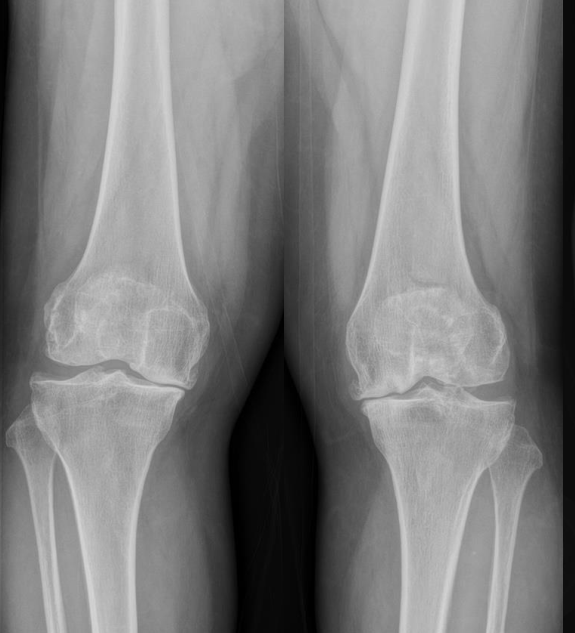
An anteroposterior radiograph of both knees from a clinical case in the lecture. It allows assessment of the joint spaces together with the morphology of the femoral and tibial articular surfaces. Professor Ro's lecture materials.
Select an image to view it at a larger size.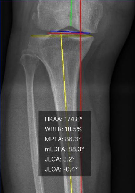
Alignment axes and joint lines are marked on a knee image from a case in the lecture. The displayed HKA, MPTA, mLDFA, and JLCA values are measurements from this case, rather than general surgical targets or acceptable limits. Professor Ro's lecture materials; CONNEVO measurement interface.
Select an image to view it at a larger size.Evidence for this topic
AAOS Guideline on Surgical Management of Knee Osteoarthritis, 2022Summary of the ESSKA European Osteotomy Consensus, 2024TKA · 02
Measured Resection and Gap Balancing
Measured resection plans bone cuts using anatomical landmarks and implant thickness. Gap balancing uses gaps measured under ligament tension after resection. In practice, surgeons often combine information from both approaches.
Wear and deformity can alter bony landmarks, while ligament condition and applied tension affect the measured gaps. Consider how the direction of the tibial cut affects femoral rotation and the flexion gap, and how additional distal femoral resection affects the joint line and mid-flexion stability.
The landmarks used in measured resection, such as the posterior condyles, transepicondylar axis, and anteroposterior axis, provide different information. Posterior condylar wear or trochlear deformity reduces the reliability of an individual landmark, so cross-check it against other references. In gap balancing, distractor tension, patellar position, knee angle, and whether osteophytes have been removed also affect the measurement. Matching gap numbers requires matching the measurement conditions first.
Resecting more distal femur solely because the extension gap is tight may elevate the joint line and affect mid-flexion stability. Flexion-gap asymmetry may arise from the tibial resection and ligament condition as well as femoral rotation. Link each additional resection, release, or component-size change to its underlying cause, then reassess the gaps and endpoints at multiple flexion angles after the change.
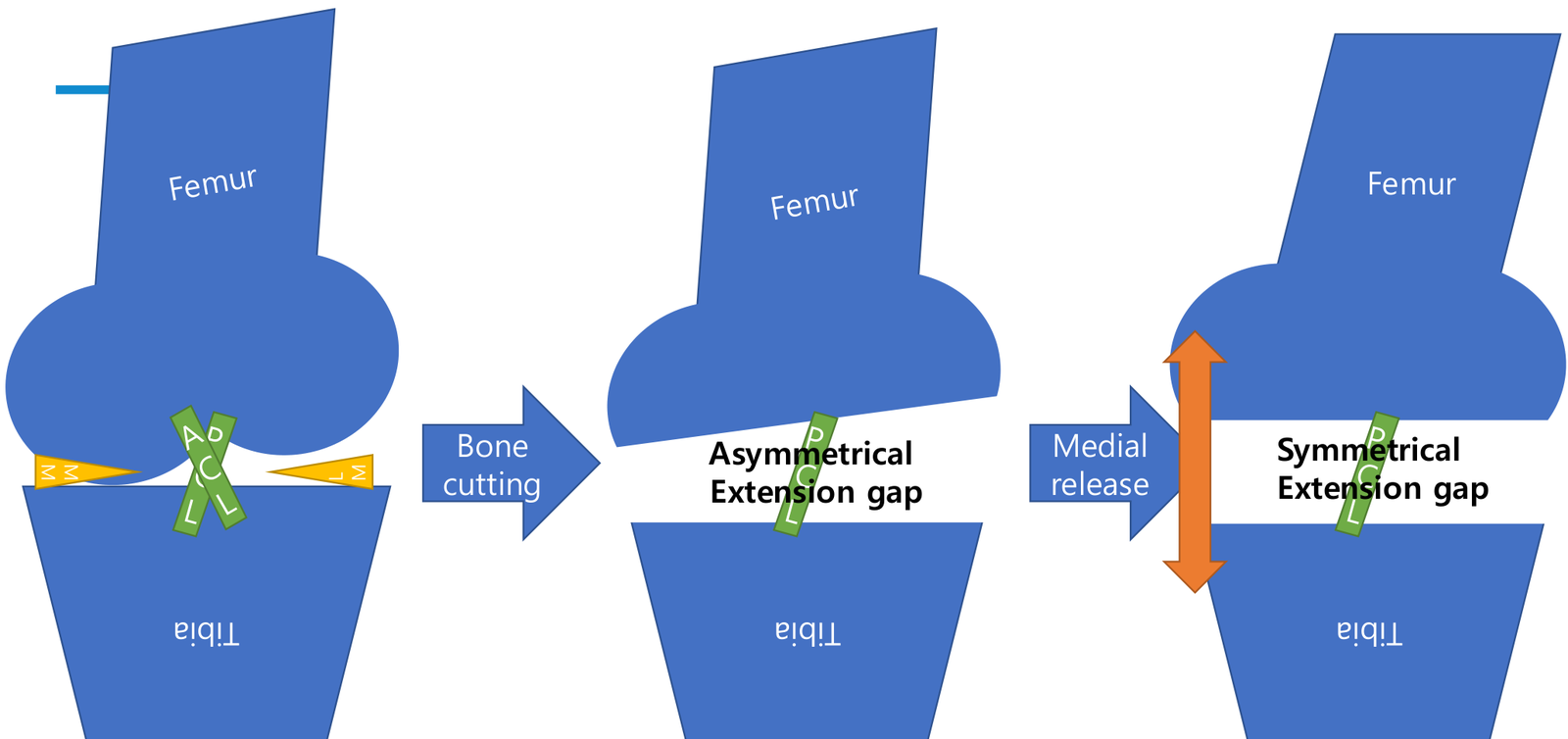
A conceptual diagram from the lecture illustrates how an asymmetric extension gap after bone cuts is corrected through medial soft-tissue release. Professor Ro's lecture materials.
Select an image to view it at a larger size.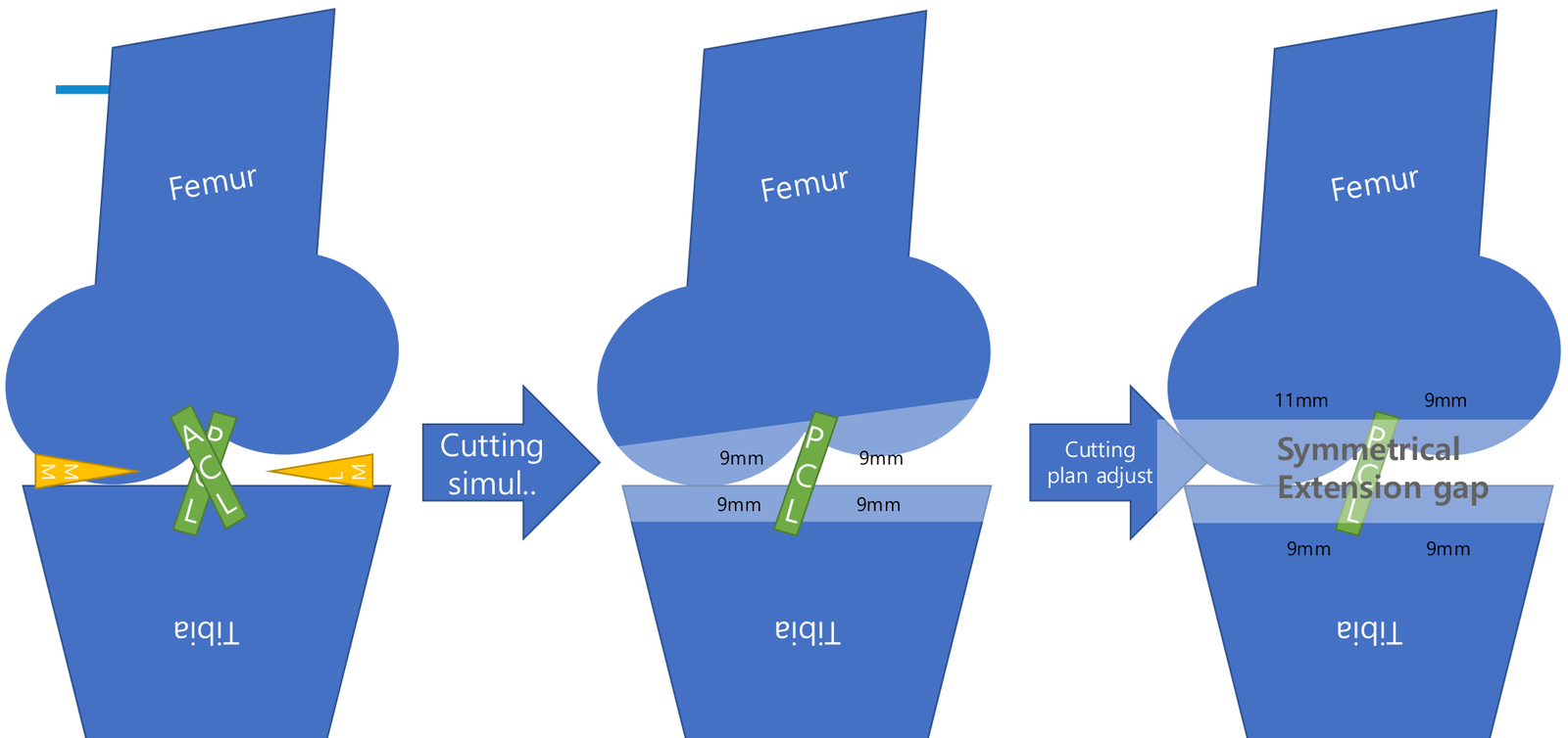
A conceptual diagram from the lecture illustrates balancing the extension gap by simulating bone cuts and then adjusting the resection plan. The displayed 9 mm and 11 mm values are examples used to explain the concept. Professor Ro's lecture materials.
Select an image to view it at a larger size.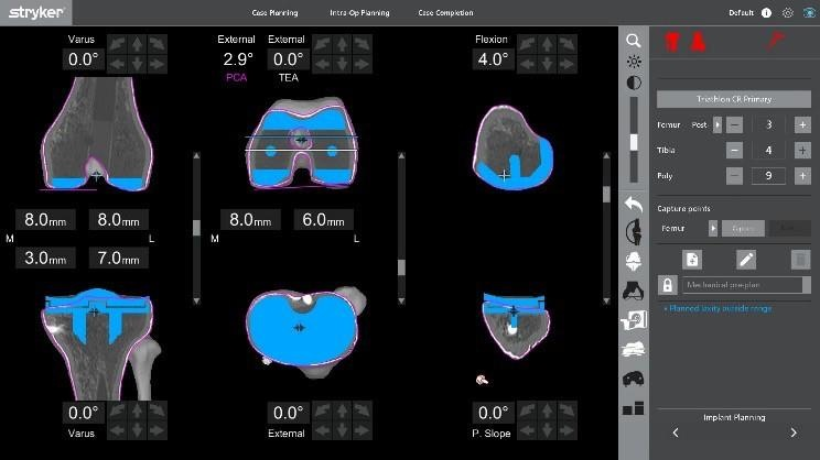
Implant planning screen for robotic surgery. The displayed angles and resection depths are examples from an individual plan, not universal targets. Screen: Stryker. Supplied by Du Hyun Ro.
Select an image to view it at a larger size.TKA · 03
Surgical Approaches and Adequate Exposure
Medial parapatellar, subvastus, and midvastus approaches differ in how they pass through the extensor mechanism and affect patellar blood supply. Skin-incision length alone does not define minimally invasive surgery. Adequate exposure is needed to assess bone cuts, rotational alignment, and ligament balance.
A limited incision can place excessive tension on structures in a knee with poor patellar mobility or a severe fixed deformity. Compare malalignment and wound problems as well as early pain and recovery. Angles and incision lengths depend on the technique and the patient, so the same values should not be applied to every knee.
Choose an approach that provides adequate exposure while preserving the extensor mechanism and patellar blood supply. Previous incisions, obesity, stiffness, severe deformity, and patella baja can make exposure more difficult and should be included in preoperative planning. Excessive patellar eversion or traction places stress on the patellar tendon insertion and soft tissues. When bone-cutting landmarks cannot be seen adequately, safe exposure takes priority over incision length.
When comparing early recovery after subvastus and midvastus approaches, assess long-term function and alignment alongside wound and extensor-mechanism complications. The label 'minimally invasive' does not guarantee implant positioning or pain outcomes. Recording why an approach was changed or extended and which structures were protected also helps assess the risks of subsequent surgery.
TKA · 04
PCL and the Flexion Gap in CR TKA
Retaining the PCL does not by itself reproduce native knee kinematics. Posterior tibial slope, posterior condylar offset, contact position, and PCL tension together influence the flexion gap. With trial components in place, check for excessive tightness or laxity at several flexion angles.
Changing polyethylene thickness to correct a gap mismatch can also alter stability at other angles. First identify whether the cause lies in resection, rotation, or the PCL. Choosing CR or PS also involves ligament function, deformity, and implant design.
In a cruciate-retaining design, the PCL influences the relationship between the femur and tibia during flexion, but its function cannot be judged simply by whether it was retained. Preoperative contracture and degeneration, injury during tibial resection, tibial slope, and the amount of posterior condylar resection alter its tension. If a trial lifts off or the contact point moves abnormally during flexion, assess component size, rotation, and the flexion gap as well as the PCL.
PCL release or sacrifice primarily changes the flexion gap, with effects that depend on its initial tension and the implant design. Increasing polyethylene thickness to reduce flexion instability may make extension excessively tight. Choosing CR, PS, ultracongruent, or constrained designs requires integrating residual ligament function, gaps, and bone loss; product names alone do not establish stability.
TKA · 05
Cemented and Biological Fixation
Cemented fixation provides immediate fixation. Cementless fixation requires initial mechanical stability followed by bone ingrowth. A porous surface has value only when sufficient initial stability is achieved. Assess cut-surface quality, bone quality, and eccentric loading together.
Failures of early cementless designs should not be grouped with outcomes of modern porous materials. Short-term results for one product do not establish long-term survival for every design. Age criteria may guide patient selection but should not be treated as indications for every patient.
Consider fixation as a problem at the load-transferring interface rather than a material label. Cemented fixation requires attention to preparation of the resected surface, cement penetration, and component seating; cementless fixation requires assessment of bone quality, press-fit, micromotion, and conditions for osseointegration. Do not equate a thin radiolucent line on a single image with loosening; relate it to pain, changes in position, and progression on serial imaging.
Failures of older cementless designs should not be generalized to all modern porous metals, and short follow-up of newer designs should not be interpreted as evidence of long-term survival. The AAOS 2022 guideline assesses fixation outcomes in light of differences between comparator groups and implant designs. When comparing studies, check femoral and tibial fixation separately, the definition of hybrid fixation, bone quality, follow-up duration, and reasons for reoperation.
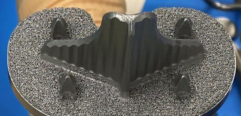
The surface of a porous tibial implant. Biological fixation requires both appropriate material and design and initial stability. Photograph: Du Hyun Ro.
Select an image to view it at a larger size.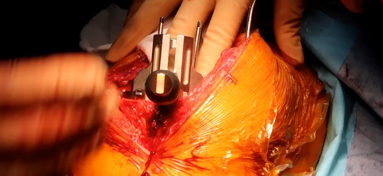
Insertion of the tibial and femoral components. Stable contact between bone and implant is assessed alongside the planned position. Photograph: Du Hyun Ro.
Select an image to view it at a larger size.Evidence for this topic
AAOS Guideline on Surgical Management of Knee Osteoarthritis, 2022TKA · 06
Mobile-bearing Design and Clinical Results
Mobile-bearing designs adjust conformity and freedom of movement to address contact stress and rotational motion. Theoretical advantages, patient-reported function, and long-term revision rates are distinct outcomes.
A moving bearing still requires appropriate bone cuts and ligament balance. A lax flexion gap or rotational problems can contribute to instability or spin-out. When comparing with fixed bearings, examine the products, indications, and follow-up periods used.
The rotational freedom of a mobile bearing is a design feature intended to address articular congruency and contact stress. Actual motion is constrained by ligament tension and component position, so a freely moving bearing is not synonymous with restoration of normal in-vivo kinematics. Rotational mobility also does not justify accepting femoral or tibial component malrotation.
When comparing designs, distinguish pain and patient-reported outcomes from wear, fixation, and reasons for revision, rather than looking at ROM alone. Evaluation of bearing dislocation or spin-out should connect gap asymmetry, excessive laxity, alignment and rotation problems, and the implant's constraint mechanism. Do not assume that a particular bearing is superior merely because a patient is active or needs deep flexion; examine the clinical results of that design.
TKA · 07
Femoral component rotation and the flexion gap
Femoral component rotation changes posterior condylar resection, the medial–lateral flexion gap relationship, and trochlear direction together. Identify the posterior condylar axis, surgical transepicondylar axis, and trochlear AP axis separately, distinguishing anatomical from surgical epicondylar axes. In a valgus knee with lateral posterior condylar hypoplasia or asymmetric wear, adding a fixed external rotation to the posterior condyles alone does not establish the target.
If bony references disagree with the tensioned flexion gap, reassess measurement conditions first. Tibial cut varus or valgus, osteophyte removal, collateral laxity, and applied tension affect gap-derived rotation. Creating a rectangular gap in an excessively lax medial compartment can induce femoral internal rotation; interpret gap shape alongside the alignment strategy and ligament condition.
When changing rotation, examine component size, AP position, and posterior condylar offset as well as medial and lateral posterior resections. Correcting asymmetry through rotation alone may worsen ligament tension at other angles or the patellofemoral relationship. With trials, assess coronal stability, contact, and patellar tracking in mid-flexion as well as extension and 90° flexion, documenting changes after further resection or release.
Do not attribute postoperative anterior pain or maltracking to rotation alone. Assess combined femoral and tibial rotation, patellar thickness and position, soft tissues, and fixation. State the reference axes and observer uncertainty in CT measurements. Angles associated with complications in clinical studies are not universal revision thresholds; symptoms, examination, and imaging should support the same mechanism.
Evidence for this topic
Femoral rotation using the epicondylar axis, 1993AP axis for femoral rotation in valgus TKA, 1995Component malrotation and patellofemoral complications, 1998TKA · 08
Tibial rotation and patellofemoral tracking
Tibial rotation is not determined solely by foot direction or maximal coverage of the cut surface. The Akagi line connects the center of the PCL attachment to the medial border of the tibial patellar tendon attachment. Consider the tibial tubercle, anterior cortex, and cut-surface morphology together, specifying each reference definition. When osteophytes, deformity, or bone loss alter a landmark, reduce reliance on it and cross-check other references.
Baseplate coverage and rotation are related but distinct objectives. Internally rotating a large tray to cover an asymmetric proximal tibia can adversely affect femorotibial rotation and patellofemoral tracking. Simply increasing external rotation may instead produce overhang and soft-tissue irritation. Adjust size, shape, and AP position together, assessing cortical support, protrusion, and planned rotation separately.
Dynamic trial alignment requires consideration of patellar position, the soft tissues after exposure, and femoral component rotation. The position into which a trial aligns during flexion and extension provides supplementary information, not a substitute for anatomical references. Check contact, impingement, bearing relationships, and patellar tracking at several angles. A mobile bearing cannot be assumed to compensate consistently for rotational mismatch.
In complex primary and revision procedures, deeper cuts can change the relationship between tibial cross-sectional morphology and reference axes. Use the same method when comparing postoperative CT rotation measurements and interpret tibial rotation alongside femoral rotation. A particular angle alone does not establish the pain mechanism or an indication for revision. Define correction goals after relating pain location and movement to fixation, infection assessment, and other possible causes.
Evidence for this topic
An anteroposterior axis of the tibia for TKA, 2004Tibial rotational references and resection level, 2010Component malrotation and patellofemoral complications, 1998TKA · 09
Joint-line height and mid-flexion instability
Joint-line height describes the position of the femoral and tibial articular surfaces relative to ligament attachments and the extensor mechanism, not merely one resection thickness. Estimate height using several landmarks, including epicondyles and the fibular head excluding osteophytes, alongside preoperative imaging and wear. Contralateral images can help, but symmetry is not assumed. Record joint-line displacement, posterior condylar offset, and patellar-height changes separately.
Additional distal femoral resection followed by a thicker insert to address an extension contracture can elevate the joint line. Cadaver experiments demonstrated increased coronal laxity in mid-flexion despite maintained extension and 90° gaps. This illustrates how moving the joint surface and rotation center relative to collateral attachments alters tension across angles. Experimental displacement values are not clinical safety limits or universal failure thresholds.
Distinguish mid-flexion instability from a globally excessive flexion gap. When stairs, sit-to-stand, or recurrent effusion cause concern, identify the angle and direction of instability and the endpoint. Ligament laxity, component position and rotation, and design can contribute alongside joint-line elevation. Acceptable static radiographs or stability in extension and at 90° do not exclude a mid-flexion problem.
During planning, separate osteophytes, posterior tissues, bone resection, and ligament condition as causes of contracture, and anticipate the consequences of further cuts. Revision joint-line restoration may require planning distal and posterior femoral defects, augments, component size, and fixation together. Before choosing a thicker insert or greater constraint, assess how it changes gaps throughout motion. Judge height restoration within the combined goals of stability, extensor function, and fixation.
Evidence for this topic
Joint-line elevation and mid-flexion laxity: cadaver study, 2018Joint-line elevation and in-vivo BCR TKA kinematics, 2021/2022Joint-line restoration and function after primary TKA, 2022TKA · 10
Prehabilitation: distinguish prognostic factors from treatment effects
An association between preoperative strength or walking ability and postoperative recovery is different from evidence that changing those factors through rehabilitation improves long-term outcomes. Prehabilitation begins with the hypothesis that improving function before surgery may support recovery, while the effects of each programme and uncertainty in the research require separate evaluation. Pain, flexion contracture, quadriceps function, chair rise and stair performance, and walking-aid use inform preparation goals for each patient.
The content and feasibility of an exercise programme matter more than its label. Assessment includes the balance of strengthening, range-of-motion work, walking and education, supervision and actual participation, pain or swelling after exercise, and limitations imposed by comorbidity. Applying the same intensity and schedule to a patient with markedly impaired function and a patient who already functions well may miss both potential benefit and burden. Goals are adapted to functional tasks the patient can perform.
In a 2022 randomized trial of 262 participants, four sessions of multidisciplinary rehabilitation and education did not significantly improve early functional independence or activity limitations over the first six months after surgery. A recent strength-training trial of 48 participants found benefits in strength and some early stair performance, but postoperative improvement in other functional measures was inconsistent. These findings neither establish that all prehabilitation is ineffective nor demonstrate long-term benefit for every patient. They concern different programmes, populations, assessment times and outcomes.
Counselling specifies realistic preoperative goals such as maintaining function within pain tolerance, practising walking-aid use and transfers, and understanding recovery. Recovery is not guaranteed, and a fixed number of exercise sessions is not presented as a requirement for surgical eligibility. Reassessment records actual participation and change in the target tasks alongside strength measurements, and connects the plan with postoperative rehabilitation and discharge support.
Evidence for this topic
Multidisciplinary prehabilitation and functional outcomes after TKA: randomized trial, 2022Preoperative maximal strength training before TKA: randomized trialTKA · 11
Fall risk across the transition from waiting to hospital and home
Falls are assessed in the distinct settings of the surgical waiting period, early hospitalization and return home. Pain, leg weakness, reduced activity and fear of falling can form a cycle that further limits movement. Recent falls and their circumstances, episodes of the knee giving way, dizziness on standing, walking-aid use and routes through the home clarify the support required at each stage.
A prospective study associated a history of preoperative falls with postoperative falling. Relief of arthritic pain therefore does not establish that fall risk has also resolved. Assessment links range of motion and strength with balance, ankle and opposite-leg function, medications and general health, and actual movement during everyday tasks. Fall rates vary with follow-up duration and study population; they are not an individual patient's probability.
Early in hospitalization, assessment includes the effects of analgesia and anaesthesia, actual quadriceps activation, getting out of bed and walking to the toilet. Findings on peripheral nerve blocks and falls differ across studies, and studies of different block sites, delivery methods and recovery pathways are not combined into one conclusion. Current motor function and supervision needs guide walking-safety assessment beyond the procedure name alone. Walking aids or immobilizers are also selected within the institutional recovery pathway and the patient's condition.
Discharge readiness extends beyond walking on level ground. Practice includes the stairs the patient will use, nighttime toilet transfers, rising from a chair, family assistance and walking-aid use, and informs the support plan. After a fall, assessment covers the wound, extensor mechanism, periprosthetic injury and systemic causes, as well as fear of movement. Documentation specifies which tasks require whose assistance rather than stopping at a list of risk factors.
Evidence for this topic
Prospective observation of falls before and after TKA, 2009TKA · 12
How back pain and other painful joints affect TKA outcomes
TKA goals distinguish pain in the operated knee from limitations in the patient's overall mobility. Back pain, other painful lower-limb joints and psychological burden can influence interpretation of outcomes. Recording pain location and radiation, and identifying limitations in standing, walking, stairs or chair rise, separates the limitations explained by the knee findings from those that suggest another source.
Degenerative changes on lumbar radiographs are not equivalent to clinically important spinal symptoms. In a study of 225 TKA patients, radiographic lumbar degeneration severity was not associated with postoperative clinical scores, whereas severe radiating pain during activity was associated with a lower postoperative physical-function score. Outcome assessment considers pain characteristics and neurological findings together with hip and ankle function and the clinical examination, beyond imaging alone.
Studies linking pain at several sites to outcomes require a distinction between univariable associations and results adjusted for other factors. Comorbidity, preoperative pain severity and emotional health can interact; a single factor has not thereby been established as the sole cause of low satisfaction. Rather than assuming that pain is exaggerated, assessment clarifies the patient's understanding of the goals, difficulties with sleep and activity, and specific barriers to participation in rehabilitation.
Surgical counselling distinguishes improvement in knee-joint pain from resolution of all painful sites and mobility limitations. Follow-up tracks knee pain, stability and range of motion alongside patient-reported outcomes and actual task performance. Slower-than-expected functional recovery prompts evaluation of knee problems such as infection, fixation failure and instability, together with reassessment of how pre-existing spine and other joint symptoms have changed.
Evidence for this topic
Lumbar degeneration, symptoms and clinical outcomes in TKA patients, 2014TKA · 13
Implant size and shape: assess AP and ML fit separately
Average differences in knee shape between populations are not a basis for selecting an individual's implant by ethnicity or sex alone. The anteroposterior (AP) length, mediolateral (ML) width and their ratio are assessed separately on the femoral and tibial resection surfaces. Measurements depend on resection level and orientation, rotation and remaining osteophytes, so measurement conditions are established on imaging and on the actual resection surface.
An AP-matched femoral component may still overhang in the ML direction. Downsizing solely to fit ML width can change the relationship to the anterior cortex, posterior condyles or flexion gap. Consideration of narrow options, component size and AP position includes anterior notching, overstuffing and posterior condylar offset as well as overhang and underhang. Improving fit in one area can change resection and soft-tissue relationships elsewhere.
In a study of 437 implantations of the same design, localized femoral component overhang was associated with clinically important pain at two years. This supports considering soft-tissue irritation; it does not establish that the study's measurement threshold is a universal implant tolerance or an indication for revision. The site of overhang is compared with tenderness, movement-related symptoms, rotation and patellofemoral relationships to judge its relevance in the individual patient.
For the tibial tray, coverage, rotation and cortical support are recorded separately. A change intended to cover more of the resection surface may alter the planned rotation or produce soft-tissue overhang. Assessment of the centre of the resection surface, deformity and bone defects precedes changes to the shape or size of a symmetric or asymmetric tray. Better morphological fit and superiority in pain, function or long-term fixation are separate outcomes.
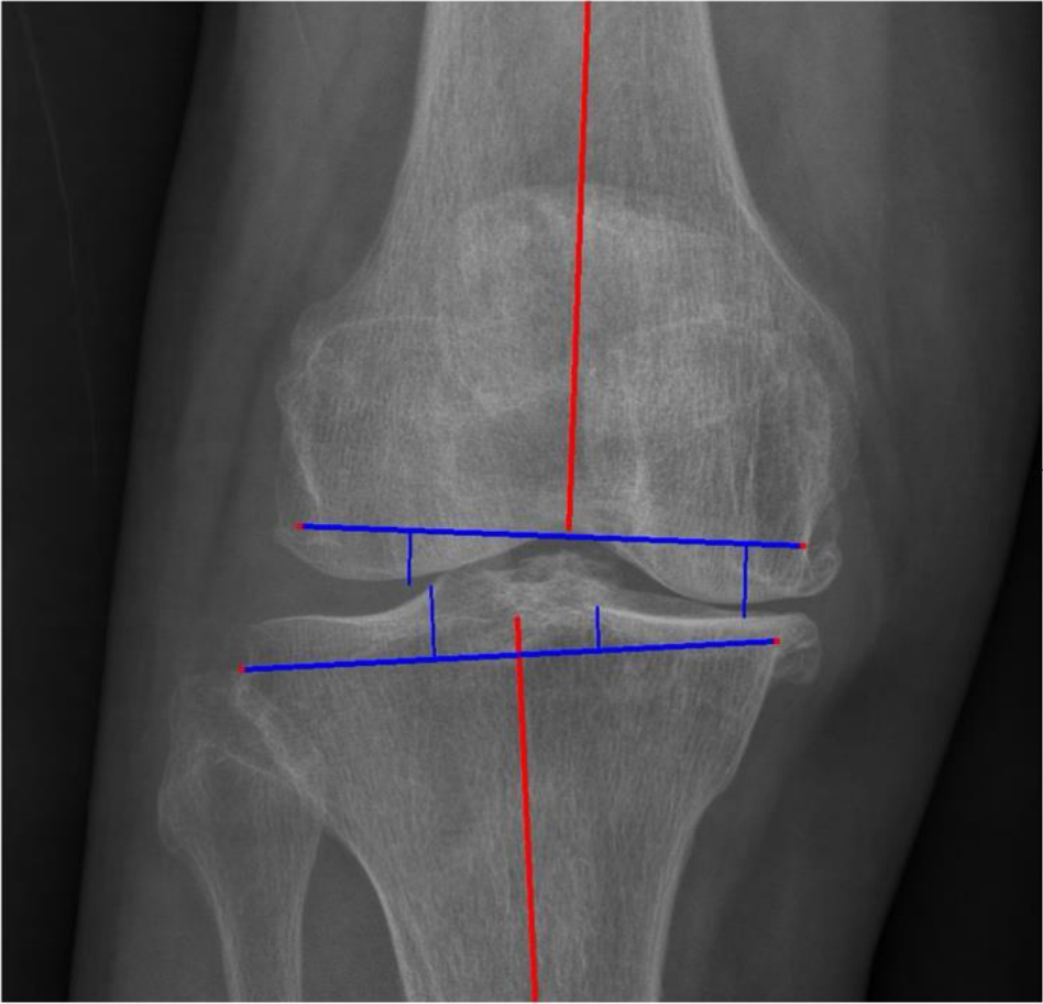
A coronal radiograph with femoral and tibial mechanical axes and planned resection lines superimposed. Cropped from Figure 1 in Park et al. (2024). CC BY 4.0.
Select an image to view it at a larger size.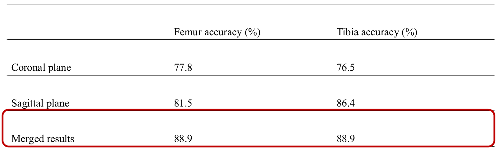
The table compares sizing agreement for coronal, sagittal, and merged predictions. Allowing a one-size margin, the merged prediction reached 88.9% for both femoral and tibial components. Park et al. (2024), lecture table by Du Hyun Ro.
Select an image to view it at a larger size.Evidence for this topic
Clinical association between femoral component overhang and pain, 2010TKA · 14
Deep flexion and floor activities: connect ROM with everyday tasks
The patient's idea of a knee that bends well is first linked to specific daily activities. Kneeling, squatting and sitting cross-legged may be insufficiently represented in general functional scores. Maximum passive flexion, a weight-bearing activity and the ability to rise from the floor are different outcomes. Alongside preoperative range of motion, assessment records how pain, strength, the opposite joint and balance limit each task.
Changes to posterior condyles, liners and post-cam mechanisms in high-flexion designs aim to address contact and movement in deep flexion. Design intent does not guarantee restoration of high-flexion activities. Resection, soft-tissue balance, posterior condylar relationships, impingement, the extensor mechanism, preoperative function and rehabilitation all affect actual movement. Changes in size or gaps to gain flexion also require assessment of stability at multiple angles and fixation.
In a randomized bilateral comparison involving 44 patients, a standard PS implant in one knee and a high-flexion concept PS implant in the other did not produce significant differences in ROM or clinical scores at two years. Another multicentre randomized trial also found similar two-year outcomes for its compared implants. These findings apply to specific products, populations and follow-up periods; they do not establish that all designs are equivalent. A small difference in maximum flexion angle and recovery of the activities the patient values remain separate outcomes.
Counselling separately records important floor activities alongside goals for pain relief and walking. Deep flexion is not promised for every patient, and an experimental interface-stress study alone does not support prohibiting all activity beyond a specific angle. Postoperative assessment establishes which tasks are possible and why others are uncomfortable, and individualizes activity and rehabilitation goals according to recovery stage, strength, balance, the implant and fixation.