REVISION · PJI · RESIDENTS & FELLOWS
Revision Arthroplasty and Complications
This section presents a structured assessment of pain and examines instability, infection, fracture, and surgical exposure for revision as distinct problems.
REVISION · PJI · 01
Instability at Different Angles and Assessment of Its Cause
Instability in extension, midflexion, and flexion does not represent one uniform problem. Connect symptoms with examination and assess component fixation, position and rotation, the joint line, and ligaments. Infection and other sources of pain must also be considered.
Polyethylene exchange or greater constraint does not solve every cause. Identify the problematic gap or structure first, then plan revision with bone defects and extensor mechanism condition in mind.
Describe instability by angle and direction, such as extension, mid-flexion, flexion, or recurvatum. Relate the history and examination to the movements that provoke giving way, difficulty on stairs, or recurrent effusion. Even when static imaging suggests acceptable positioning, ligament competence and dynamic gaps may differ; assess component fixation and rotation, the joint line, and posterior condylar relationships together.
Isolated polyethylene exchange changes insert height but does not directly correct problems in resection, rotation, or joint-line position. Increased constraint likewise requires consideration of ligament function, bone defects, and fixation demands. After assessing other causes of pain, including infection, plan the target gaps and any necessary bone reconstruction, stems, or constraint. Distinguish an improvement in stability from correction of its underlying cause.
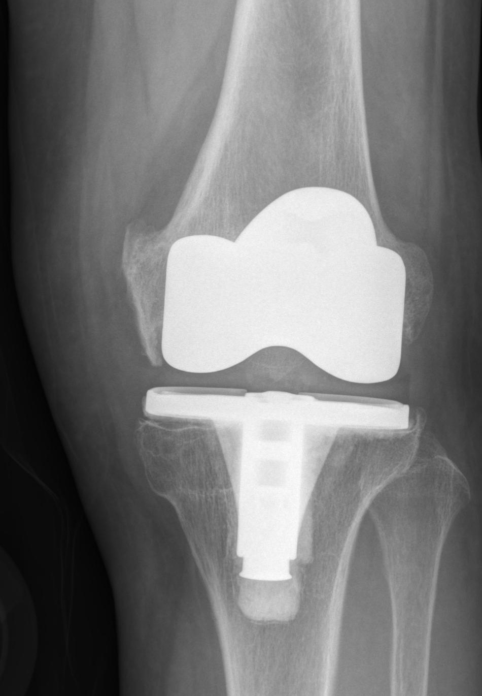
Pre-revision anteroposterior radiograph from a case of wear and aseptic loosening.
Select an image to view it at a larger size.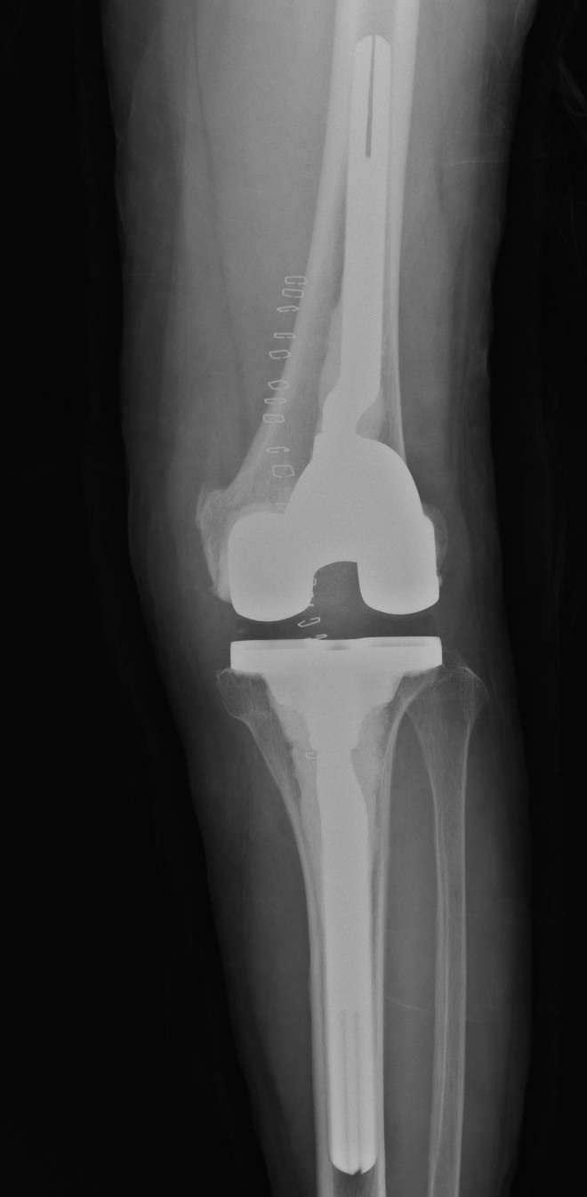
Post-revision anteroposterior radiograph showing stemmed femoral and tibial components.
Select an image to view it at a larger size.REVISION · PJI · 02
Combining Test Results in PJI Assessment
Assess PJI using history, wound condition, symptoms, inflammatory markers, and synovial-fluid and microbiological results together. Time since surgery and prior antibiotic use affect interpretation. One negative test does not exclude infection.
The acute or chronic course, implant fixation, soft tissues, causative organism, and the patient’s general condition influence treatment strategy. Interpret infection test results using current consensus definitions and institutional clinical standards. Antibiotic choice and dosage take culture results, antimicrobial susceptibility, and the patient’s general condition into account.
PJI assessment begins with the presence of a sinus tract, wound history, timing of pain, systemic symptoms, and previous antibiotics. Serum markers, synovial cell count and differential, cultures, and histology provide different information, and interpretation changes in the early postoperative period, inflammatory disease, or metallosis. Do not equate a negative culture with absence of infection; examine sampling conditions and consistency among microbiological and other test results.
The EBJIS definition expresses uncertainty through the categories infection unlikely, likely, and confirmed. Meeting diagnostic criteria does not in itself establish an indication for DAIR or staged revision. Discuss treatment in relation to symptom duration, organism and susceptibility, implant fixation, soft tissues, and patient condition. Plan with specialist collaboration and institutional protocols, and do not treat a diagnostic checklist as a substitute for antibiotic prescribing.
Evidence for this topic
EBJIS Definition of Periprosthetic Joint Infection, 2021REVISION · PJI · 03
Key Decisions in Periprosthetic Fractures
Alongside fracture location and displacement, assess whether the implant is fixed and how much bone remains. Also consider the extensor mechanism, soft tissue, and what surgery and rehabilitation the patient can tolerate.
Fractures around a fixed implant and those around a loose implant need different plans. Classification organizes information but does not establish whether fixation or revision is feasible. Discuss bone quality, union, and failure risks together.
In a periprosthetic fracture, assess component stability and residual bone stock as well as fracture location, pattern, and displacement. Pain before the fracture and serial images provide clues to preexisting loosening. The femoral component's box and stem affect whether a retrograde nail can be used; existing hardware and stems in other joints should also be included when planning the whole construct and avoiding stress risers.
Fixation may be considered for a fracture around a stable implant, and revision reconstruction for loosening with substantial bone loss, but the actual choice also reflects bone quality, soft tissues, and patient function. A patellar fracture additionally requires assessment of extensor-mechanism continuity. When proposing early weight-bearing, explain whether the procedure, fixation stability, and associated lesions can accommodate that load.
REVISION · PJI · 04
Revision Exposure and Protection of the Extensor Mechanism
Scarring, stiffness, previous incisions, and patellar height affect revision exposure. Obtain an adequate view while protecting skin perfusion and continuity of the patellar and quadriceps tendons.
Quadriceps snip, V–Y approaches, and tibial tubercle osteotomy expand exposure differently. Compare the required view, extensor function, bone quality, fixation, and healing. Understand the risk of forceful traction through limited exposure.
Plan revision exposure beginning with skin perfusion, previous incisions, and soft-tissue condition. Multiple incisions, scarring, stiffness, and patella baja increase wound and extensor-disruption risks as well as technical difficulty. Consider the necessary extension of the approach instead of placing excessive traction on the patellar tendon in a restricted field, and obtain sufficient visualization for component removal and reconstruction of bone defects.
Quadriceps snip, V–Y approaches, and tibial tubercle osteotomy differ in the exposure obtained and the remaining functional and healing conditions. Incorporate the TTO fragment, fixation, and union, and tension and strength after V–Y, into planning; do not rank techniques by their names alone. Postoperative exercise and loading instructions must relate to how the extensor mechanism was handled. The operative description of exposure is important for later rehabilitation and subsequent surgery.
REVISION · PJI · 05
Bone loss and fixation strategy in revision arthroplasty
In revision arthroplasty, focus on how the remaining bone supports loading and fixation rather than defect size alone. Alongside AORI classification, record location, containment, cortical walls, and collateral-ligament attachments separately for the femur and tibia. Plain radiographs can underestimate defects hidden by implants, and removal can cause further bone loss. Include infection assessment and previous operative records, and prepare for both anticipated and actual intraoperative defects.
Consider cement, bone graft, or metal augments for small focal defects and cones, sleeves, or structural reconstruction for major metaphyseal defects. Relate selection to residual bone quality, surrounding walls, and implant support area rather than fixed depth thresholds. Additional resection intended to eliminate a defect can sacrifice joint-line position and bone stock. First determine the joint line, femoral rotation, and posterior condylar relationships to restore, then select augments and metaphyseal support accordingly.
The zonal concept divides fixation into epiphyseal bone near the joint surface, metaphysis, and diaphysis. A stem transfers load and supplements stability but does not automatically restore the joint line or component position. If canal direction restricts the desired position, assess offsets and stem fixation options. Cones and sleeves differ in coupling and preparation and are not interchangeable labels; consider insertion fracture, fixation, and the difficulty of later removal.
Long-term series of porous metaphyseal support report low aseptic-loosening rates, but reoperation for infection or instability still occurs. Observational comparisons have not consistently demonstrated that adding a cone or sleeve reduces every cause of re-revision. Connect bone reconstruction with constraint selection appropriate to ligament function. During follow-up, assess component-specific fixation, progressive radiolucencies, pain, and infection signs. Stable imaging and success of the whole knee are not equivalent.
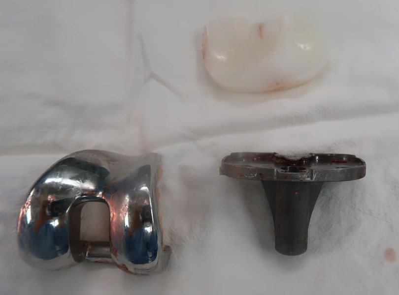
Femoral and tibial components and a polyethylene insert removed during revision surgery.
Select an image to view it at a larger size.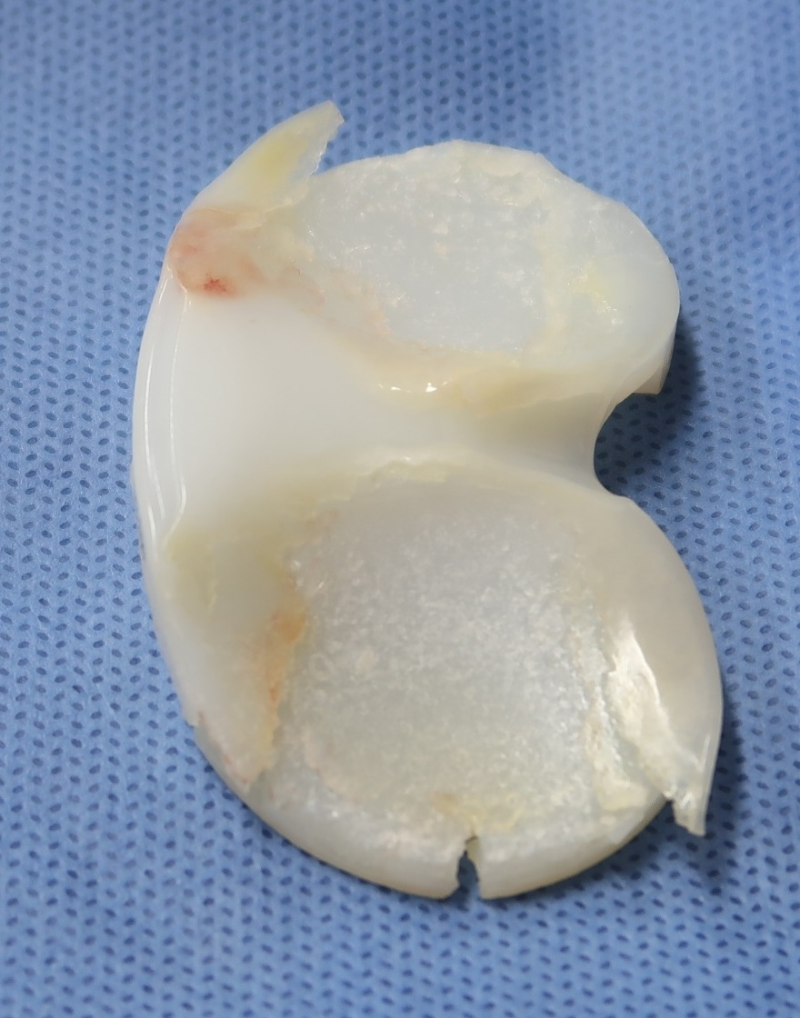
The articular surface of a retrieved polyethylene insert shows surface damage and edge defects.
Select an image to view it at a larger size.Evidence for this topic
International consensus on management of bone loss in revision arthroplasty, 2024Long-term follow-up of porous tibial metaphyseal cones, 2024Registry comparison of constructs adding cones or sleeves to stems, 2025REVISION · PJI · 06
Constraint selection and joint stability
In revision arthroplasty, constraint determines how much motion the implant must restrict when damaged ligaments cannot provide restraint. First record the direction, flexion angle, and endpoint of instability, and assess bone loss, ligament attachments, and the extensor mechanism. If flexion–extension mismatch arises from component position or joint-line errors, correct recoverable causes before increasing constraint. Distinguish ligamentous instability from giving way associated with infection, loosening, or pain.
A PS design substitutes for part of PCL function but does not address major collateral-ligament incompetence. CCK or varus–valgus constrained designs use interaction between a larger post and box to limit coronal and rotational motion in a nonlinked construct. The permitted motion varies by design; identical labels do not imply identical constraint. Assess residual collateral function and the direction of laxity remaining at different angles with trials in place to select the required design.
A rotating hinge mechanically links the femur and tibia while permitting axial rotation. It may be considered for extensive ligament deficiency or severe instability, but permitted rotation does not eliminate load at the bone–implant interface. Stress distributions in CCK and hinge designs depend on linkage, stems, and activity conditions and cannot be predicted from a simple linear ranking of constraint. Plan metaphyseal support, stems, and component position together.
Some clinical comparisons of revision patients found similar long-term function and survival with CCK and rotating-hinge designs, but differences in patient selection and implant design limit generalization. Select a design that provides adequate stability; insisting on insufficient constraint can leave residual instability. With final trials, check gaps at several angles, dislocation potential, and extensor-mechanism movement. Follow stability together with fixation, pain, infection, and component-related problems.
Evidence for this topic
Long-term comparison of CCK and rotating hinge in revision arthroplasty for extension instability, 2022Numerical study of stress and micromotion in CCK and rotating-hinge designs, 2019Comparison of implant-interface stress under collateral-ligament deficiency, 2018REVISION · PJI · 07
Patellar fracture after TKA: extensor function, fixation and remaining bone
After TKA, patellar fracture treatment cannot be determined from displacement alone as in a simple traumatic fracture. Three axes guide assessment: continuity of the extensor mechanism, fixation of the patellar component and remaining bone stock. Acute trauma and an incidentally detected chronic fracture are distinguished. Review includes pain onset, active extension and extensor lag, skin and wound condition, previous resurfacing and revision records together with imaging.
The Ortiguera–Berry classification defines type I as an intact extensor mechanism with a stable component, type II as disruption of the extensor mechanism, and type III as a loose component with an intact extensor mechanism. Within type III, remaining bone stock further informs the feasibility of reconstruction. Documentation includes actual active-extension function and the evidence for fixation alongside the classification label. Uncertainty in extension testing due to pain and effusion is not proof of normal function.
In the original clinical series, nonoperative results were generally favorable in type I fractures with preserved fixation and extensor function, whereas operated cases with extensor disruption or component loosening had substantial complications and reoperations. These findings challenge the assumption that every fracture requires surgery. Protection, activity modification and follow-up reflect acuity, displacement, symptoms and extensor function; a historical immobilization duration is not a universal schedule regardless of patient condition.
When surgery is needed, preparation accounts for thin or poorly vascularized remaining patella, fragment size, defects after component removal and the feasibility of extensor mechanism reconstruction. Successful bone union and recovery of active extension, pain and function are separate outcomes. Assessment also includes factors such as malrotation or tracking problems that may worsen loading. Treatment choices are explained in relation to the structures that can be preserved, expected function, infection and nonunion risk, and the patient's activity requirements.
Evidence for this topic
Classification and treatment outcomes of patellar fractures after TKA, 2002REVISION · PJI · 08
Two-stage PJI treatment: reimplantation timing requires more than one number
In two-stage exchange for infection, the interval after spacer insertion is an ongoing treatment process leading up to reimplantation. Prereimplantation ESR and CRP, spacer exchanges and time to reimplantation have been studied in relation to treatment failure. The initial organism and specimen reliability, antimicrobial treatment, wound healing, drainage, pain, systemic condition and mechanical spacer problems are recorded chronologically to clarify the basis for the decision.
A decrease or normalization of ESR and CRP does not by itself confirm eradication of infection. In a multicentre analysis of 291 patients, percentage changes in ESR and CRP between resection and reimplantation lacked sufficient diagnostic ability to distinguish reinfection risk. Absolute values and trends are considered with the clinical course, and the spacer status and whether antimicrobials were being administered or withheld at the time of testing are recorded. A single result does not automatically invalidate other reasons for clinical suspicion.
A retrospective study of 101 patients found a higher failure risk when reimplantation occurred more than 18 weeks after explantation, but this is not a universal reimplantation deadline. Persistent infection, health status and additional treatment may contribute to a longer interval. An observed association is different from a treatment effect of earlier reimplantation itself. Interpretation of interval-based results includes the definition of failure and the handling of patients who never reached reimplantation or underwent spacer exchange.
Reimplantation planning integrates clinical recovery and available test results with the judgment of infectious-disease and reconstruction teams. Preparation accounts for expected bone defects, soft tissues and the extensor mechanism, spacer-related changes, and definitive fixation and constraint. Patient counselling explains why one laboratory value cannot determine the date and which issues still need to be resolved. Outcomes are evaluated through reinfection, repeat procedures, eventual reimplantation and functional recovery together.