Symptoms and tests
Do all painful knees need an MRI?
First ask which treatment decision the test could change.

At a glance
- Select tests to answer questions from symptoms and examination.
- An MRI finding is not automatically the pain source or a reason for surgery.
- Ask how the result could change treatment.
MRI provides detailed images, but it need not be the first test for every painful knee. Tests are selected to answer questions raised by symptoms and examination. Having more images is not the same as understanding the cause better.
Three questions when choosing a test
- 01
Clinical question
Which tissue or problem is suspected?
- 02
Suitable test
Choose the information needed from examination, X-ray or MRI.
- 03
Treatment decision
How would the result change exercise, medicine or surgery planning?
Three panels connect the clinical question, test information and treatment decision
Questions answered by examination and X-rays
The clinician asks about onset, location, injury, swelling and instability and examines movement. Typical osteoarthritis may be diagnosed from symptoms and examination; imaging is not essential in every case.
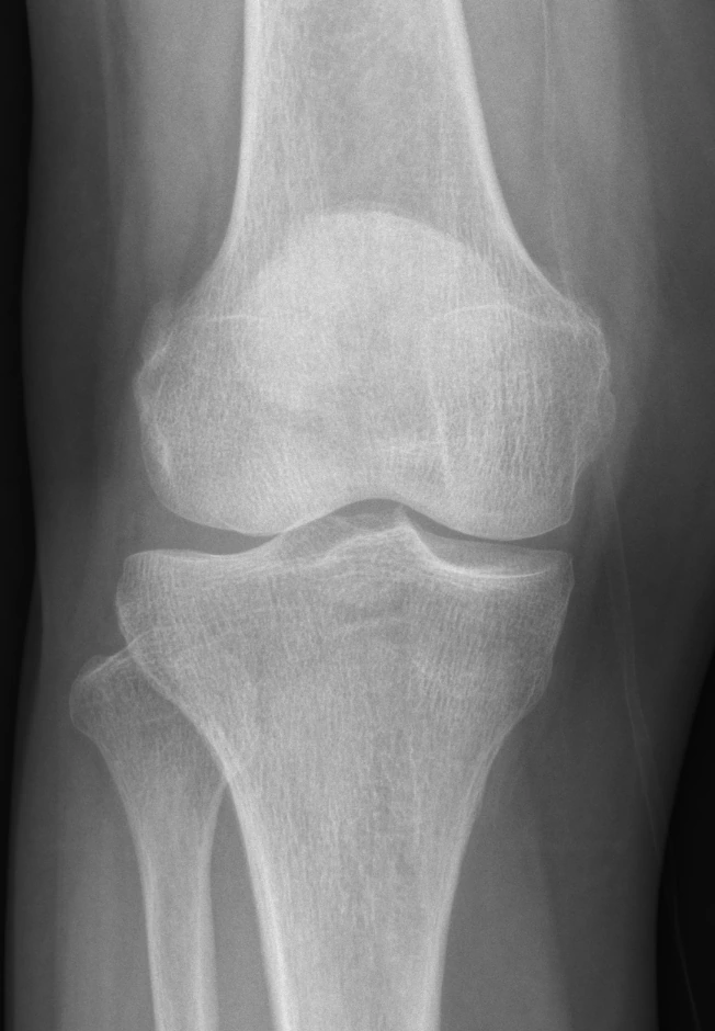
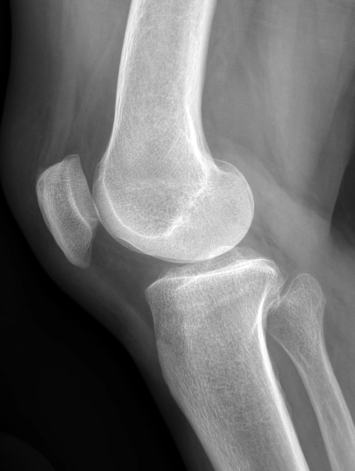
X-rays assess bones, joint space, spurs and alignment. Weight-bearing and patellofemoral views may be chosen for different questions. When surgery is considered, identifying the extent of structural change and alignment is an important imaging role.
Some questions benefit from MRI
MRI can show menisci, ligaments, articular cartilage and changes inside bone in more detail. It may help when ligament or meniscal injury is suspected after trauma, when there is true locking, or when symptoms and existing tests do not fit well.
Ask which tissue needs assessment and how the result could alter treatment. Repeat MRI is not always needed to follow the ordinary course of known arthritis. New symptoms, examination, earlier images and the treatment plan guide the decision.
Connect report terms to symptoms
A report mentioning degeneration or a meniscal tear does not establish that it is the main cause of pain. Structural changes may be found without symptoms. Ask how the location of a finding matches examination rather than focusing only on unfamiliar words.
One image or report should not determine surgery. Associated arthritis, instability, actual functional limitation and response to nonsurgical care matter too. The word tear does not automatically mean tissue must be removed; preservation and rehabilitation should also be considered.
Clarify purpose and safety before the test
Ask what the test aims to find out, whether its result will change treatment, and whether another test could provide the information. If you have had previous tests, give their dates and bring the images and reports if possible. This helps determine whether repeating a test is necessary.
Tell the imaging team in advance about a pacemaker or other implanted device, previous surgery involving metal, possible pregnancy, or difficulty with enclosed spaces. MRI does not use radiation, but the type of device and your condition can affect whether it is suitable and what preparation is needed.
Symptoms and examination define the question for imaging
At the visit, we first ask when the pain began, whether there was an injury, which activities worsen it, and whether there is swelling or catching. We also review previous treatments, other health conditions, and current medicines. Examination assesses gait, knee movement, tenderness, swelling, and stability to identify findings that relate to the symptoms.
Typical osteoarthritis can be assessed through the history and examination, so imaging is not required for everyone. When an X-ray is needed, it shows structural features such as the joint space, bone spurs, and changes in bone shape. The results are interpreted alongside the symptoms and examination.
MRI provides detailed images of soft tissues such as the menisci and ligaments. It can be useful, for example, when an acute meniscal injury is suspected after trauma. The decision to use MRI considers the question being investigated and how the findings could help guide treatment.
Common Questions
Is MRI always better than an X-ray?
They provide different information. Bone, weight-bearing alignment and soft tissues require different assessment approaches.
Should arthritis be followed with regular MRI?
Not routinely when there is no new problem or question relevant to management.
Does a tear in the report mean surgery?
Symptoms, examination, tear pattern, associated disease and treatment response guide the decision.
Evidence and references
01When to seek medical care
Each warning sign is a separate reason to seek care. Follow the action below if any one applies.
Emergency department · call 119 in Korea
- Call 119 for sudden breathlessness or chest pain, severe faintness or loss of consciousness. Do not wait for calf swelling to appear.
- Go to an emergency department if you suddenly cannot bear any weight, or if an injured knee swells rapidly or looks deformed. A cold or numb foot after injury also needs immediate assessment.
- Seek emergency care for a hot, swollen and severely painful joint with high fever, shaking chills, confusion or inability to bear weight.
- With back or leg pain, go immediately to an emergency department for new difficulty passing urine, loss of bladder or bowel control, or numbness around the genitals or anus. Severe or rapidly worsening weakness or numbness in both legs also needs immediate assessment.
Same-day assessment
- Arrange assessment today for a newly hot, swollen joint, sudden severe pain, or a knee locked so that it cannot fully straighten. Joint infection can occur without fever.
- Have new swelling or pain in one calf assessed today for a possible blood clot. Call 119 immediately if breathlessness or chest pain develops.
- Severe knee pain or a new limp in a growing adolescent may come from a hip condition such as slipped capital femoral epiphysis. Stop walking and sport and arrange assessment today. Go directly to emergency care if they cannot bear weight.
- Stop running and seek assessment today for new recurring groin pain, pain with walking or at night, or a limp. A femoral neck stress fracture needs to be considered. Keep weight off the painful leg until assessed. Go to emergency care if you cannot bear weight.
Book an outpatient appointment
- If none of the warning signs above is present, book an appointment for pain lasting a few weeks, repeated swelling or reduced daily activity and exercise.