2025 · Orthopaedic Journal of Sports Medicine
Can Landmark-based PTS Be Measured on Limited-field Lateral Radiographs or Images without a Calibration Scale?
ICCs against manual measurement were 0.91 for the short axis and 0.92 for the long axis. Automated repeat-measurement ICC was 1.00.
Results explained with study numbers
 Enlarge Figure
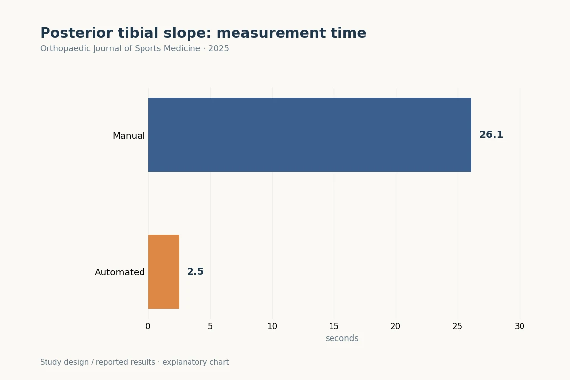
Manual measurement took 26.1 seconds and automated measurement took 2.5 seconds.
Orthopaedic Journal of Sports Medicine (2025). DOI: 10.1177/23259671251331067 · Original explanatory chart based on reported data
The research question
Participants and Methods
Main Findings
Implications for clinical and surgical practice
Limitations
Let's put it briefly.
Which clinical question does this address?
This is a study of standardizing PTS measurement. Clinical cutoffs should not be transferred unchanged between different shaft-axis protocols.
Keywords
- automated measurement
- computer vision model
- deep learning
- landmark-based
- posterior tibial slope
- AI
- ACL
- HTO
Abstract · Original text
ByeongYeong Ryu, Jun Woo Nam, Du Hyun Ro, R Kyle Martin, Eivind Inderhaug, Andreas Persson, Sanna Haland, Sung Eun Kim. Automated Posterior Tibial Slope Measurement Using Lateral Knee Radiographs: A Novel Landmark-Based Approach Using Deep Learning. Orthopaedic journal of sports medicine. 2025;13(4):23259671251331067. doi:10.1177/23259671251331067.
BACKGROUND
A single universally accepted protocol does not exist for measuring the posterior tibial slope (PTS), limiting the application of cutoff values for surgical decision-making and risk stratification.
PURPOSE/HYPOTHESIS
This purpose of this study was to validate an online computer vision model using anatomic landmarks for PTS measurement on uncalibrated lateral knee radiographs. It was hypothesized that this model would achieve similar accuracy to manual measurement.
STUDY DESIGN
Cohort study; Level of evidence, 2.
METHODS
A total of 10,007 lateral knee radiographs collected between January 2009 and December 2019 were utilized. The data set comprised 9277 (93%) training, 500 (5%) validation, and 230 (2%) test radiographs. After defining "A" as the distance from the tibial joint line to the proximal aspect of the tibial tuberosity, 2 landmark-based methods for determining the tibial shaft axis were developed based on lines connecting the tibia midpoints at distances: (1) 2A and 3A (short method) and (2) 2A and 4A (long method). The PTS was then calculated using each tibial shaft axis. Model performance was evaluated against orthopaedic specialists' measurements using inter- and intraobserver intraclass correlation coefficients (ICCs). Model performance on shortened images, subcategorized into normal, osteoarthritic, and implant-embedded knees, was also assessed, along with time efficiency comparisons.
RESULTS
The overall interobservers ICCs were 0.91 for the short method and 0.92 for the long method against manual measurement. The ICCs for normal, osteoarthritic, and implant-embedded radiographs were 0.84, 0.90, and 0.97 for the short method and 0.88, 0.91, and 0.97 for the long method, respectively. The intraobserver ICC for the computer vision model was a perfect 1.00, while manual measurements showed ICCs of 0.89 for the short method and 0.95 for the long method. The mean model measurement time was 2.5 ± 0.7 seconds, compared with 26.1 ± 1.9 seconds for the manual measurement (P < .001).
CONCLUSION
A novel, time-efficient, deep learning model for measuring PTS demonstrated excellent accuracy and consistency across various lateral knee radiographs. If externally validated, this model may enable a pathway for direct clinical translation of research findings by providing a standardized measurement tool.
© 2025 The Author(s)
The original paper.
Automated Posterior Tibial Slope Measurement Using Lateral Knee Radiographs: A Novel Landmark-Based Approach Using Deep Learning
ByeongYeong Ryu, Jun Woo Nam, Du Hyun Ro, R Kyle Martin, Eivind Inderhaug, Andreas Persson, Sanna Haland, Sung Eun Kim
Orthopaedic Journal of Sports Medicine · 2025