R.노두현정형외과와 연구 이야기
글자
← 연구 이야기

무릎 모양과 변화 · 2025

관절염이 심한 무릎은 모양도 다를까요?

관절염 등급이 다른 무릎 17,365개에서 정렬 유형의 분포를 비교한 연구입니다.

Journal of Bone and Joint Surgery American Volume · Sung Eun Kim, Samuel MacDessi, Daeseok Song, Joong Il Kim, Byung Sun Choi, Hyuk-Soo Han, Du Hyun Ro

연구 질문과 설계

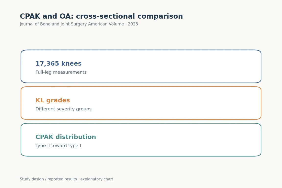
17,365개 무릎에서 KL 등급별 CPAK·aHKA·JLO 분포를 비교했습니다.그림 크게 보기

17,365개 무릎에서 KL 등급별 CPAK·aHKA·JLO 분포를 비교했습니다.

Journal of Bone and Joint Surgery American Volume (2025). DOI: 10.2106/JBJS.24.00316 · Original explanatory chart based on reported data

근거·원문그림 이용조건
01

관절염 등급과 다리 모양을 함께 비교했습니다

같은 관절염이라는 이름 안에도 무릎의 형태는 여러 가지입니다. 저희는 17,365무릎의 전장 X-ray를 자동 측정해 관절염 KL 등급과 CPAK 정렬 유형을 함께 살폈습니다. 관절염이 없는 집단과 심한 집단에서 형태가 어떻게 다른지, 나이의 차이만으로 설명되는지를 묻는 연구입니다.

02

그림에서 가장 흔한 유형이 달라집니다

낮은 KL 등급에서는 CPAK II형이, 높은 등급에서는 I형이 더 흔한 양상이 관찰됐습니다. 관련된 각도도 서로 다른 방향으로 변했습니다. 허벅지뼈와 정강이뼈의 관절면 각도, 두 관절선 사이 각도를 나누어 읽어야 O자 다리라는 한 표현으로 놓치는 정보를 확인할 수 있습니다.

03

나이만의 문제로 설명하기는 어려웠습니다

나이를 나누어 분석해도 관절염 중증도와 형태의 관련성이 중요했습니다. 오래 살았기 때문에 같은 모양으로 변한다고 단순하게 설명할 수는 없습니다. 원래 형태가 관절염에 취약했을 가능성과 관절염 과정에서 형태가 달라졌을 가능성이 모두 남아 있습니다. 어느 쪽이 먼저인지에 대해서는 추적 연구가 필요합니다.

04

한 사람의 변화 과정을 그린 그림은 아닙니다

이 연구는 서로 다른 무릎들을 같은 시점의 정보로 비교한 횡단면 연구입니다. 그림에서 II형의 비율이 줄고 I형이 늘어도, 한 환자가 반드시 II형에서 I형으로 변한다는 뜻은 아닙니다. 여러 집단의 차이를 개인의 진행 경로로 바꾸어 읽지 않는 것이 가장 중요한 제한입니다.

05

수술 계획에는 현재 사진과 원래 형태를 함께 봅니다

인공관절 수술을 연구할 때 현재의 변형을 그대로 원래 모양이라고 생각해도 되는지가 중요합니다. 이 논문은 그 질문을 더 구체적으로 만듭니다. 다만 어떤 정렬 철학이 더 좋은 수술 결과를 내는지는 별도의 비교 연구가 확인해야 합니다. 분류 이름만으로 본인의 수술 방법을 정하지는 않습니다.

연구 숫자 다시 보기

17,365분석한 무릎
KL 등급관절염 정도 구분
CPAK정렬 형태 분류
RESEARCH FINDING무릎

연구 대상의 구성

분석한 무릎
17,365
09,98519,970
연구에 포함된 자료의 수입니다. 치료의 효과나 개인의 위험 확률을 나타내지 않습니다.

많이 묻는 질문

이 결과를 개인의 치료에 바로 적용할 수 있나요?

인공관절 수술을 연구할 때 현재의 변형을 그대로 원래 모양이라고 생각해도 되는지가 중요합니다. 이 논문은 그 질문을 더 구체적으로 만듭니다. 다만 어떤 정렬 철학이 더 좋은 수술 결과를 내는지는 별도의 비교 연구가 확인해야 합니다. 분류 이름만으로 본인의 수술 방법을 정하지는 않습니다.

연구 방법과 수치 자세히 보기

Coronal Plane Alignment of the Knee (CPAK) Type Shifts Toward Constitutional Varus with Increasing Kellgren and Lawrence Grade: A Radiographic Analysis of 17,365 Knees

대상과 방법

17,365 knees. 심한 골 소실 >5 mm, Ahlbäck 4–5, 이전 골 수술 등을 제외했습니다.

AI 영상 측정에 기반한 후향적 단면연구입니다. KL·CPAK 유형과 연령에 따라 정렬 지표를 비교했습니다.

논문에 보고된 결과

  1. KL이 증가할수록 최빈 CPAK은 II형에서 I형으로 이동했습니다(P < 0.05). LDFA·JLCA는 증가했고 KL 2 이후 HKAA·MPTA·aHKA는 감소했습니다.
  2. 연령과 LDFA·MPTA의 연관성은 제한적이었습니다.

해석의 범위

개별 환자의 종단적 CPAK 변화를 입증한 것은 아닙니다. 본래 내반의 OA 취약성과 OA에 의한 해부학적 변화를 구별할 수 없습니다.

Keywords · 주제어

Abstract · 원문 초록

Sung Eun Kim, Samuel MacDessi, Daeseok Song, Joong Il Kim, Byung Sun Choi, Hyuk-Soo Han, Du Hyun Ro. Coronal Plane Alignment of the Knee (CPAK) Type Shifts Toward Constitutional Varus with Increasing Kellgren and Lawrence Grade: A Radiographic Analysis of 17,365 Knees. The Journal of bone and joint surgery. American volume. 2025;107(3):297-303. doi:10.2106/JBJS.24.00316.

BACKGROUND

Studies investigating constitutional alignment across various grades of osteoarthritis (OA) are limited. This study explored the distribution of Coronal Plane Alignment of the Knee (CPAK) types and associated radiographic parameters with increasing OA severity.

METHODS

In this retrospective cross-sectional study, 17,365 knees were analyzed using deep learning software for radiographic measurements. Knees were categorized on the basis of the Kellgren and Lawrence (KL) grade and CPAK type. Radiographic measurements were the hip-knee-ankle angle (HKAA), lateral distal femoral angle (LDFA), medial proximal tibial angle (MPTA), arithmetic HKAA (aHKA), joint line obliquity (JLO), and joint line convergence angle (JLCA). Age-stratified analysis was performed to differentiate the impact of age on OA severity.

RESULTS

A shift in the most common CPAK type from II to I was found with increasing KL grade (p < 0.05). Furthermore, there was a corresponding increase in LDFA and JLCA with increasing KL grade, while HKAA, MPTA, and aHKA decreased after KL grade 2. Age exhibited limited association with LDFA and MPTA, suggesting that OA severity is the dominant factor related to the CPAK distribution.

CONCLUSIONS

The study found a shift in CPAK type with worsening OA. It is possible that constitutional varus types are more susceptible to OA, or that their increased OA prevalence is related to anatomical changes. This analysis offers new insights into alterations in CPAK type that occur with OA and underscores the importance of understanding pre-arthritic anatomy when performing joint reconstruction.

LEVEL OF EVIDENCE

Prognostic Level III . See Instructions for Authors for a complete description of levels of evidence.

Copyright © 2024 The Author(s). Published by The Journal of Bone and Joint Surgery, Incorporated.