Sung Eun Kim, Samuel MacDessi, Daeseok Song, Joong Il Kim, Byung Sun Choi, Hyuk-Soo Han, Du Hyun Ro. Coronal Plane Alignment of the Knee (CPAK) Type Shifts Toward Constitutional Varus with Increasing Kellgren and Lawrence Grade: A Radiographic Analysis of 17,365 Knees. The Journal of bone and joint surgery. American volume. 2025;107(3):297-303. doi:10.2106/JBJS.24.00316.
BACKGROUND
Studies investigating constitutional alignment across various grades of osteoarthritis (OA) are limited. This study explored the distribution of Coronal Plane Alignment of the Knee (CPAK) types and associated radiographic parameters with increasing OA severity.
METHODS
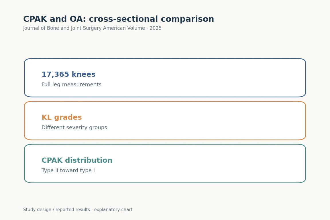
In this retrospective cross-sectional study, 17,365 knees were analyzed using deep learning software for radiographic measurements. Knees were categorized on the basis of the Kellgren and Lawrence (KL) grade and CPAK type. Radiographic measurements were the hip-knee-ankle angle (HKAA), lateral distal femoral angle (LDFA), medial proximal tibial angle (MPTA), arithmetic HKAA (aHKA), joint line obliquity (JLO), and joint line convergence angle (JLCA). Age-stratified analysis was performed to differentiate the impact of age on OA severity.
RESULTS
A shift in the most common CPAK type from II to I was found with increasing KL grade (p < 0.05). Furthermore, there was a corresponding increase in LDFA and JLCA with increasing KL grade, while HKAA, MPTA, and aHKA decreased after KL grade 2. Age exhibited limited association with LDFA and MPTA, suggesting that OA severity is the dominant factor related to the CPAK distribution.
CONCLUSIONS
The study found a shift in CPAK type with worsening OA. It is possible that constitutional varus types are more susceptible to OA, or that their increased OA prevalence is related to anatomical changes. This analysis offers new insights into alterations in CPAK type that occur with OA and underscores the importance of understanding pre-arthritic anatomy when performing joint reconstruction.
LEVEL OF EVIDENCE
Prognostic Level III . See Instructions for Authors for a complete description of levels of evidence.
Copyright © 2024 The Author(s). Published by The Journal of Bone and Joint Surgery, Incorporated.