2025 Korean Knee Society Continuing Education Course
Diagnosis and Treatment of Wear, Osteolysis, and Aseptic Loosening After Total Knee Arthroplasty
This lecture examines wear-particle generation and the biological response that produces osteolysis after total knee arthroplasty, then considers surveillance, insert exchange, and revision based on infection assessment and component fixation.
Select an image to view it at a larger size.
The Clinical Relationship Between Wear, Osteolysis, and Loosening
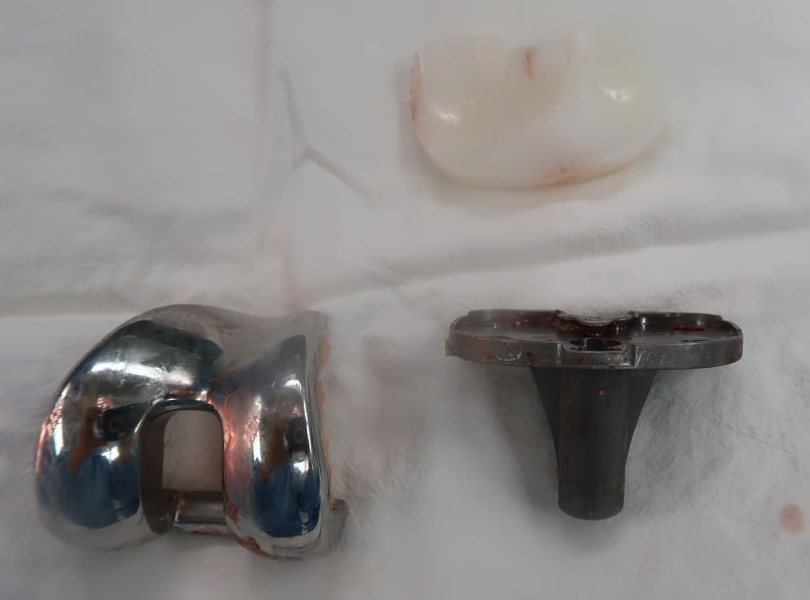
Long-term follow-up after total knee arthroplasty requires consideration of polyethylene wear, osteolysis, and aseptic loosening. Wear is the process in which material damage at the articular surface or insert backside generates particles. Osteolysis is the loss of periprosthetic bone through a biological response to those particles, whereas loosening means that a component no longer maintains mechanical fixation. These processes are related, but the presence of osteolysis does not mean that every component is already loose.
Aseptic loosening can arise from particle-induced osteolysis, but also from failure of initial fixation or micromotion at the fixation interface. When evaluating a painful knee arthroplasty, symptoms alone do not establish the failure mechanism. Component position and fixation, bone loss, and instability must be assessed together. Infection can also cause pain, effusion, and bone loss, so its possibility must be evaluated before attributing failure to an aseptic cause.
Materials and Mechanics That Influence Polyethylene Wear
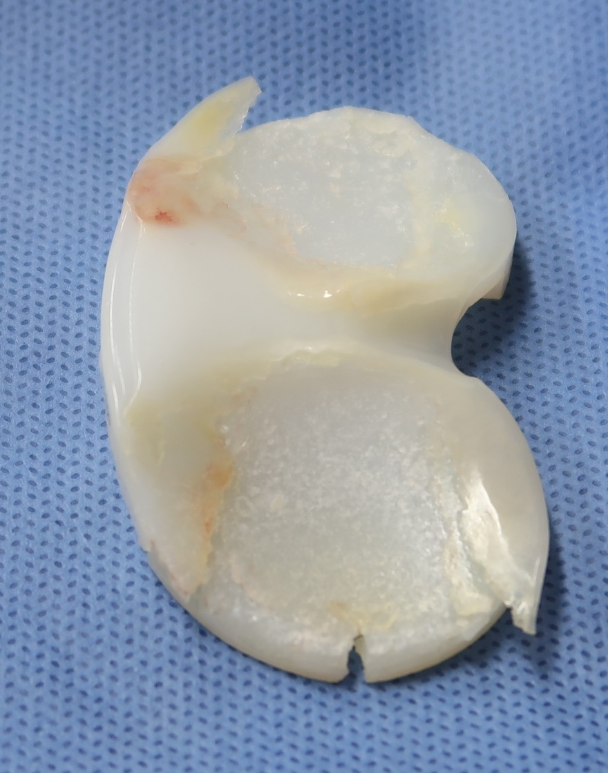
Repeated loading and friction can produce scratching, pitting, delamination, and permanent deformation of polyethylene. Because the knee combines rolling, sliding, and rotation, contact patterns and kinematics influence the form of wear. Articular geometry, component alignment, and soft-tissue balance can concentrate loading on one region or produce edge loading when they are unfavorable. Third-body material, such as cement trapped between bearing surfaces, can also increase wear.
Sterilization and processing also matter. Residual free radicals after irradiation can promote oxidation, while crosslinking and thermal treatment affect the balance among wear resistance, oxidative stability, and fatigue strength. Stabilization with vitamin E is one approach to improving these material properties. In modular tibial components, micromotion between the insert backside and metal tray can also generate particles, making the locking mechanism and tray surface relevant. A material designation or fixation method alone does not establish the long-term superiority of an individual implant.
The Biological Pathway From Wear Particles to Osteolysis
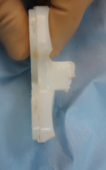
As wear particles accumulate in the joint and periprosthetic tissues, macrophages engulf them and initiate an inflammatory response. Mediators such as TNF-α and IL-1β promote osteoclast formation and activity, increasing bone resorption. Tissue responses depend on particle size, properties, and routes of migration as well as the amount of debris. Severe wear can allow metal-to-metal contact, generating metal particles that may also contribute to the periprosthetic response.
Progressive osteolysis reduces the bone supporting a component, weakens its fixation interface, and increases micromotion. That motion can contribute to further particle generation and dissemination, worsening bone loss and mechanical instability. Osteolysis can nevertheless cause few symptoms while components remain well fixed. Follow-up therefore considers the progression of bone defects and component fixation over time, alongside the presence or absence of pain.
Infection Assessment and Imaging
Assessment includes the postoperative course, pain pattern, recurrent effusion, and instability during activity. Periprosthetic joint infection must also be considered when osteolysis or loosening is suspected. Blood tests such as ESR and CRP can aid infection assessment, while synovial leukocyte count, neutrophil percentage, and cultures may be obtained when clinically indicated. Infection and aseptic loosening are distinguished by integrating the clinical findings rather than relying on a single symptom or test result.
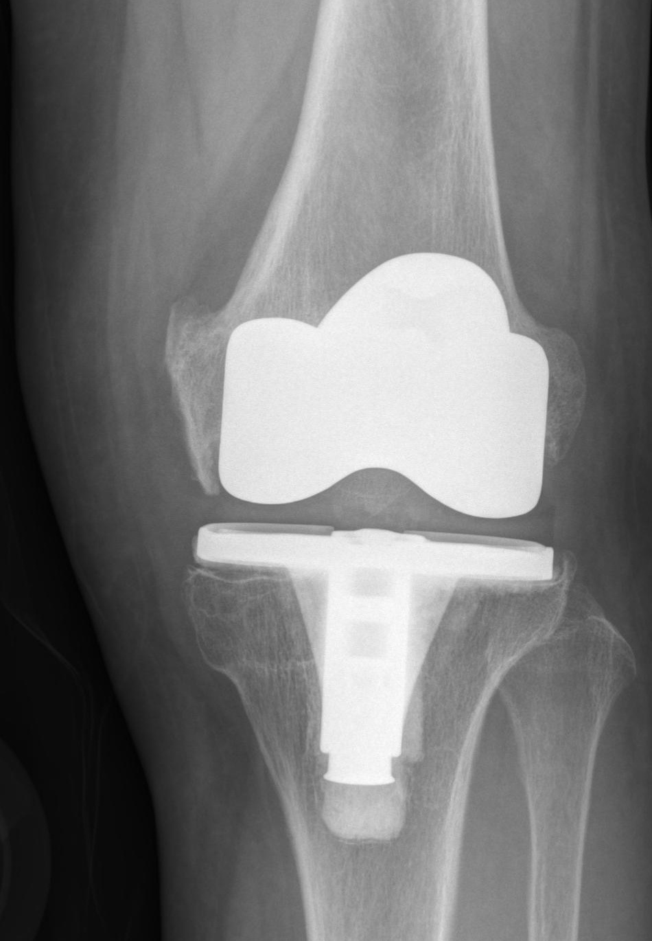
Plain radiographs are compared with earlier studies to assess progressive radiolucent lines, focal osteolysis, component subsidence, and changes in position. The Knee Society radiographic system helps document fixation interfaces and the location of bone defects consistently by zone. Metal components can obscure defects and make them appear smaller on radiographs. CT with metal artifact reduction may therefore be considered when the extent is uncertain or revision is planned. MRI with metal artifact reduction techniques can also assess marrow, synovium, and other surrounding tissues when needed.
Treatment and Revision Planning Based on Fixation
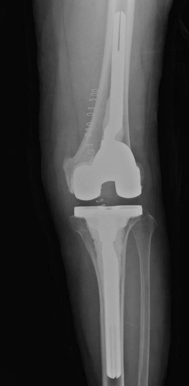
Treatment decisions integrate symptoms, the pattern of osteolysis progression, and component fixation. When components remain stable and the patient is asymptomatic, serial radiographs can monitor changes in bone defects. In selected patients with well-fixed components and wear as the principal problem, lesion curettage, bone grafting, and polyethylene insert exchange may be considered. This choice requires assessment of whether existing component fixation and joint stability can be maintained after insert exchange.
Definite component loosening or extensive bone loss calls for revision planning. Intraoperative defects may exceed the extent anticipated on imaging, so the remaining bone after component removal and the structural support required should be considered in advance. Depending on the location and size of the defects, preparation can include bone graft, metal augments and cones, and stemmed components, with stability matched to ligament function. Drugs targeting the biological pathway of osteolysis remain under investigation and are not considered substitutes for surgery addressing progressive wear and mechanical failure.
Related papers
- Goodman and Gallo: Periprosthetic Osteolysis: Mechanisms, Prevention and Treatment (2019)
- Meneghini et al.: Development of a Modern Knee Society Radiographic Evaluation System (2015)
- Haider et al.: Material Testing of Vitamin E-Stabilized Polyethylene for Total Knee Arthroplasty (2012)
- AAOS: Diagnosis and Prevention of Periprosthetic Joint Infections Clinical Practice Guideline (2019)