Korea University Hospital Continuing Education for Practicing Physicians
The Changing Alignment Paradigm in Total Knee Arthroplasty
This lecture expands the discussion from neutral alignment as a shared goal to the selection of targets tailored to each patient's anatomy and soft tissues. It explains the principles of patient-specific alignment planning by connecting constitutional varus and CPAK, arthritic deformity, the evidence and limitations of functional alignment, and AI-based measurement and prediction.
Select an image to view it at a larger size.
From Neutral Alignment to Patient-Specific Targets
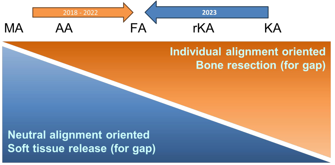
In total knee arthroplasty, mechanical alignment (MA) aims to restore a neutral mechanical axis of the lower limb and position the femoral and tibial components in the coronal plane perpendicular to their respective mechanical axes. Extensive clinical experience has accumulated with this strategy, which reproduces a standardized target and manages load transfer through the components. Understanding these basic principles is the starting point for learning newer strategies. Alignment accuracy and the patient's perception of knee function, however, are different concepts. Satisfaction also depends on pain, expectations, soft-tissue condition, physical function, and multiple surgical factors.
The question of alignment is expanding from "How accurately was the target angle achieved?" to "What target is appropriate for this patient?" Even lower limbs with the same neutral alignment can differ in femoral and tibial morphology, joint line obliquity, and medial and lateral ligament tension. Surgical planning therefore requires an integrated interpretation of the joint line and soft tissues, rather than reliance on the HKA (hip–knee–ankle angle) alone. The precise bone cuts provided by robotics or navigation are tools for implementing this judgment; they do not establish the validity of the chosen target.
MA, anatomical alignment (AA), kinematic alignment (KA), restricted kinematic alignment (rKA), and functional alignment (FA) have different starting points and adjustment principles. KA seeks to restore the pre-arthritic articular surfaces and native anatomy, while rKA follows this approach within defined limits for component and limb alignment. FA incorporates both the patient's anatomy and the soft-tissue balance assessed during surgery. Understanding this spectrum should not lead to the uniform application of one strategy to every patient.
Describing Native Alignment: Constitutional Varus and CPAK
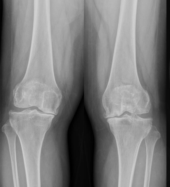
Healthy knees are not all neutrally aligned. The study of constitutional varus by Bellemans and colleagues demonstrated that natural varus alignment is also present in asymptomatic young adults. This concept helps us avoid automatically equating an angle outside neutral alignment with pathological deformity. At the same time, the existence of natural varus does not establish the safety of every degree or form of postoperative varus. Targets that respect the patient's anatomy must be considered alongside the long-term stability of the components.
CPAK (Coronal Plane Alignment of the Knee) is a classification that describes coronal alignment along two axes. aHKA (arithmetic HKA), calculated as MPTA minus LDFA, describes bony lower-limb alignment, while JLO (joint line obliquity), calculated as the sum of these two angles, describes the orientation of the joint line. Their combinations define nine phenotypes. Patients with similar HKA values can belong to different phenotypes if the femoral and tibial contributions or the joint line orientation differ. CPAK provides a shared language for describing these differences in surgical planning and research.
A phenotype classification is not a prescription that automatically assigns an alignment strategy. Even after identifying the CPAK type, ligament stability, flexion contracture, bone defects, the severity of arthritis, and intraoperative gaps must still be assessed. The distribution of normal alignment can also vary by sex, age, and population, so the average of one population should not be treated as the correct target for an individual patient. Both knees and previous images can inform the assessment, while recognizing that the contralateral knee may also have arthritis or deformity.
Distinguishing Current Deformity from Pre-Arthritic Alignment
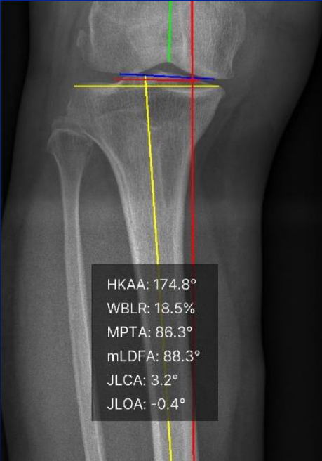
Preoperative images of patients with end-stage arthritis may not faithfully represent the native knee. Cartilage loss and bone erosion alter the height and shape of the articular surfaces, while ligament laxity or contracture changes the relative positions of the femoral and tibial articular surfaces. MPTA and LDFA, which describe bony alignment, must therefore be interpreted separately from JLCA (joint line convergence angle), which describes convergence between the two articular surfaces. It is important to determine how much of the varus reflected in the current HKA arises from bone morphology and how much arises from the joint space and soft tissues.
In a radiographic analysis of 17,365 knees in which we participated, the distribution of CPAK types differed with increasing Kellgren–Lawrence (K–L) grade, with the most common type shifting from II to I. This raises concerns about equating the phenotype measured in advanced arthritis with pre-arthritic anatomy. The study was cross-sectional, however. Comparisons of groups at different stages of arthritis cannot establish an individual patient's previous type or show that every patient underwent the same change in type.
In a longitudinal analysis of 10,841 lower limbs published in 2026, a larger JLCA and a higher baseline K–L grade, among other factors, were associated with faster changes in coronal alignment. These findings indicate that alignment should be evaluated as a process of change over time, rather than as an angle at a single time point. The observed associations, however, do not provide a basis for predicting an individual patient's progression with certainty. Changes on serial imaging, symptoms, function, and clinical examination must be considered together when deciding how much of the current deformity to correct and which anatomical features to preserve.
Functional Alignment: Incorporating Soft-Tissue Information into Bone-Cut Planning
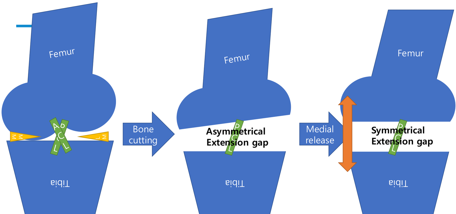
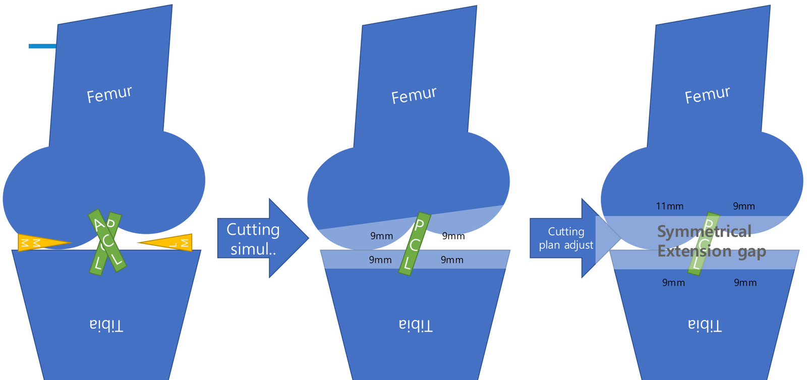
In a traditional gap-balancing approach, medial–lateral gap differences after predetermined bone cuts are addressed through soft-tissue release. FA incorporates ligament tension and extension and flexion gap information assessed before bone cuts or during surgery into decisions about component positioning. Gaps are adjusted by considering rotation and sagittal positioning as well as coronal positioning, with the aim of reducing unnecessary soft-tissue release where feasible. The resection depths and gaps shown in the figures are conceptual examples illustrating these differences, rather than resection depths to be applied directly to an actual patient.
The core of FA is neither unconditional restoration of the native joint line nor gap balancing alone. Limb alignment, component positioning, joint line preservation, and soft-tissue balance must all fall within acceptable ranges. These ranges and the sequence of adjustments vary with the surgical protocol, component design, and patient condition, making it difficult to define a particular angle as a universal criterion for FA. Even with precise measurement tools, knees with markedly impaired ligament function or severe bone defects require a separate assessment of stability and a reconstruction plan.
The 2023 study by Choi and colleagues compared robotic-assisted FA with manual MA and reported differences in patient-reported outcomes at two years. Because this was a retrospective matched study in which both the alignment strategy and the surgical tools differed, the results cannot readily be attributed to FA alone. In the 2025 randomized study by Young and colleagues comparing FA and MA within robotic-assisted surgery, there was no significant difference in the overall Forgotten Joint Score at two years, while differences were found in the frequency of soft-tissue release, some outcomes, and phenotype subgroups. The potential of FA should be considered in the context of study design, outcome measures, and patient populations, rather than taken as proof of universal superiority or better long-term implant survival.
From AI Measurement to Prediction and Patient-Specific Planning
AI-based image analysis can help standardize repeated alignment measurements and organize HKA, MPTA, LDFA, JLCA, and CPAK phenotypes in a consistent format. The purpose at this stage is to support comprehensive interpretation and comparison of the information needed from imaging. Automated measurement errors can arise from patient positioning, image coverage and quality, or anatomical deformity, so physicians must verify that the landmarks and axes correspond to the actual anatomy. Automatically calculated values do not in themselves define appropriate surgical targets.
Radiomics and time-series image analysis seek prognostic information in image texture, morphology, and structural changes in addition to alignment angles. They may contribute to future models that predict arthritis progression, functional recovery, and patient-reported outcomes. Associations between imaging features and outcomes, the performance of a prediction model, and the benefit of applying that model to actual treatment decisions are separate questions requiring validation. Until reproducibility in external populations and prospective clinical evaluation have been established, AI should not be described as automatically determining the optimal alignment strategy.
In clinical practice, the overall lower-limb axis is assessed on weight-bearing full-length radiographs, and the CPAK phenotype is interpreted by distinguishing femoral and tibial alignment from the joint line. JLCA, arthritis severity, changes on previous imaging, and clinical examination are then considered together to assess the relationship between current deformity and native anatomy. During surgery, the plan is adjusted according to the extension and flexion gaps and soft-tissue function, and the chosen strategy and reasons for adjustments are documented. The changing alignment paradigm involves more than replacing one number with another. It requires the selection of a target that can be explained by considering the patient's anatomy and function, the trajectory of change, and the level of evidence together.
Related papers
- Bellemans et al. Constitutional varus: Is neutral alignment normal for all patients? CORR, 2012
- MacDessi et al. Coronal Plane Alignment of the Knee (CPAK) classification. Bone & Joint Journal, 2021
- Kim, MacDessi, Ro et al. CPAK distribution by K–L grade: An analysis of 17,365 knees. JBJS, 2025
- Choi, Ro et al. Patient-reported outcomes of robotic-assisted functional alignment and manual mechanical alignment. KSSTA, 2023
- Young et al. A randomized comparison of functional and mechanical alignment. Journal of Arthroplasty, 2025
- Kang, Ro et al. Association between JLCA and alignment progression: A longitudinal analysis of 10,841 lower limbs. KSSTA, 2026