Hallym University Hospital Symposium
Personalized Planning for Total Knee Arthroplasty Using AI
Personalized planning for total knee arthroplasty begins with accurate measurement of each patient’s anatomy and connects implant size, alignment, and position to the surgical procedure. This lecture examines the roles and limitations of AI-assisted 2D and 3D templating, the use of risk and recovery prediction, and the validation required in clinical workflows.
Select an image to view it at a larger size.
Accurate Planning Starts with Objective Measurement
Personalized planning starts by quantifying lower-limb alignment and bone morphology using consistent definitions. On a full-length radiograph, the hip-knee-ankle angle (HKA) is assessed using the axis from the femoral head center to the distal femoral center and the axis from the proximal tibial center to the ankle center. Reviewing the joint line, femoral and tibial morphology, and bone defects connects component sizing and resection planning to anatomical evidence.
Radiographs project a three-dimensional structure onto two dimensions, so limb rotation, flexion contracture, and anterior femoral bowing can change the measurements. Values displayed to decimal precision remain susceptible to positioning and landmark errors. In Heller and colleagues’ retrospective study of 100 patients, agreement was high for some linear measurements, while reproducibility was low for certain angular measurements, including the joint line convergence angle. Imaging conditions and magnification calibration should be standardized, and automated axes and joint lines checked against the anatomy.
2D Templating: Planning Size and Alignment Together
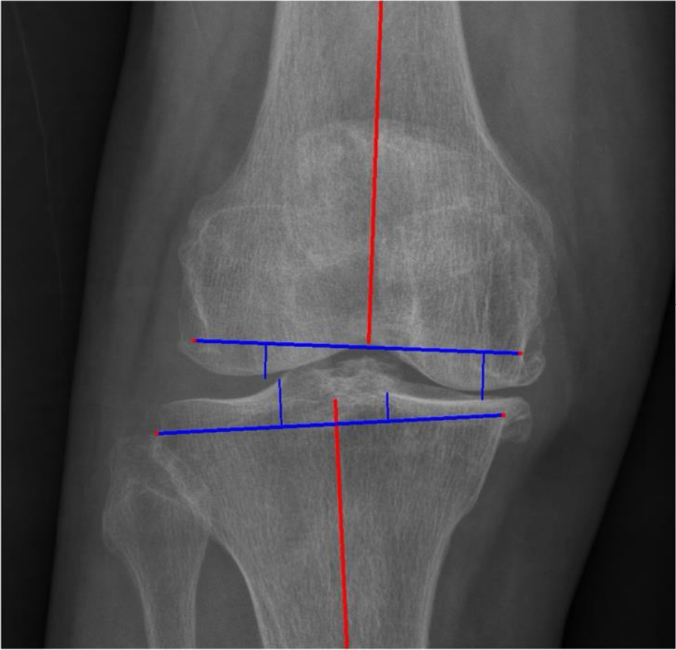
AI-assisted 2D templating uses calibrated anteroposterior and lateral radiographs to identify bone contours and landmarks and calculate component size and position. The AP view assesses mediolateral width and coronal alignment, while the lateral view assesses anteroposterior length and sagittal positioning. Our model selects the smaller size predicted from the two views to limit overhang and applies resection rules based on mechanical alignment. Interpreting an automated plan therefore requires checking the software’s alignment targets and resection rules.
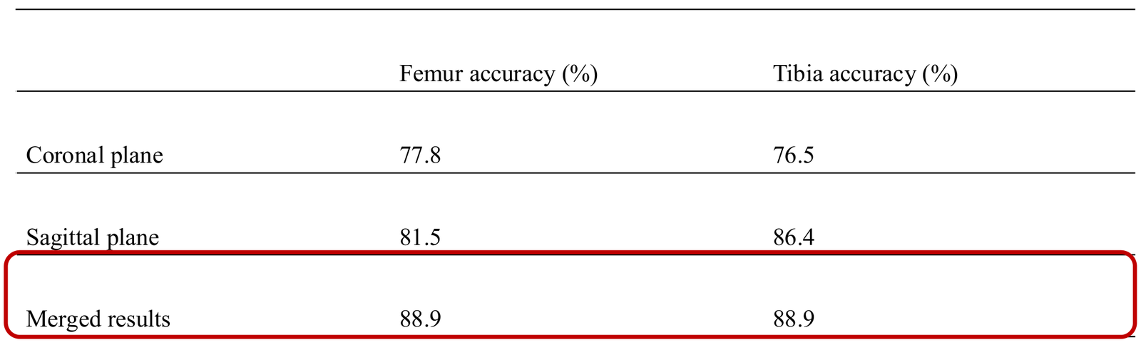
Validation in 81 procedures performed in 72 patients at Seoul National University Hospital found that 88.9% of femoral and tibial size predictions were within one size of the implanted component. This measure must be distinguished from an exact size match. On the same dataset, the specialist’s predictions were within one size in 95.1% of femoral and 100% of tibial components; measured planning time was 48.7 seconds for AI and 97.5 seconds for manual templating. These results concern the Triathlon system at a single institution and should not be assumed to apply unchanged to other institutions or implant systems.
Sizing and position or angle prediction should also be evaluated separately. In this study, comparison with postoperative radiographs showed mean positional differences of approximately 3–4 mm, while the mean sagittal femoral alignment difference was 5.1°. A size prediction within the permitted range still requires review of the anterior cortex, overhang, resection planes, and rotational positioning.
Connecting 3D Planning with Intraoperative Verification
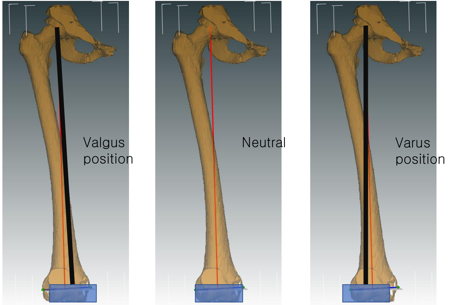
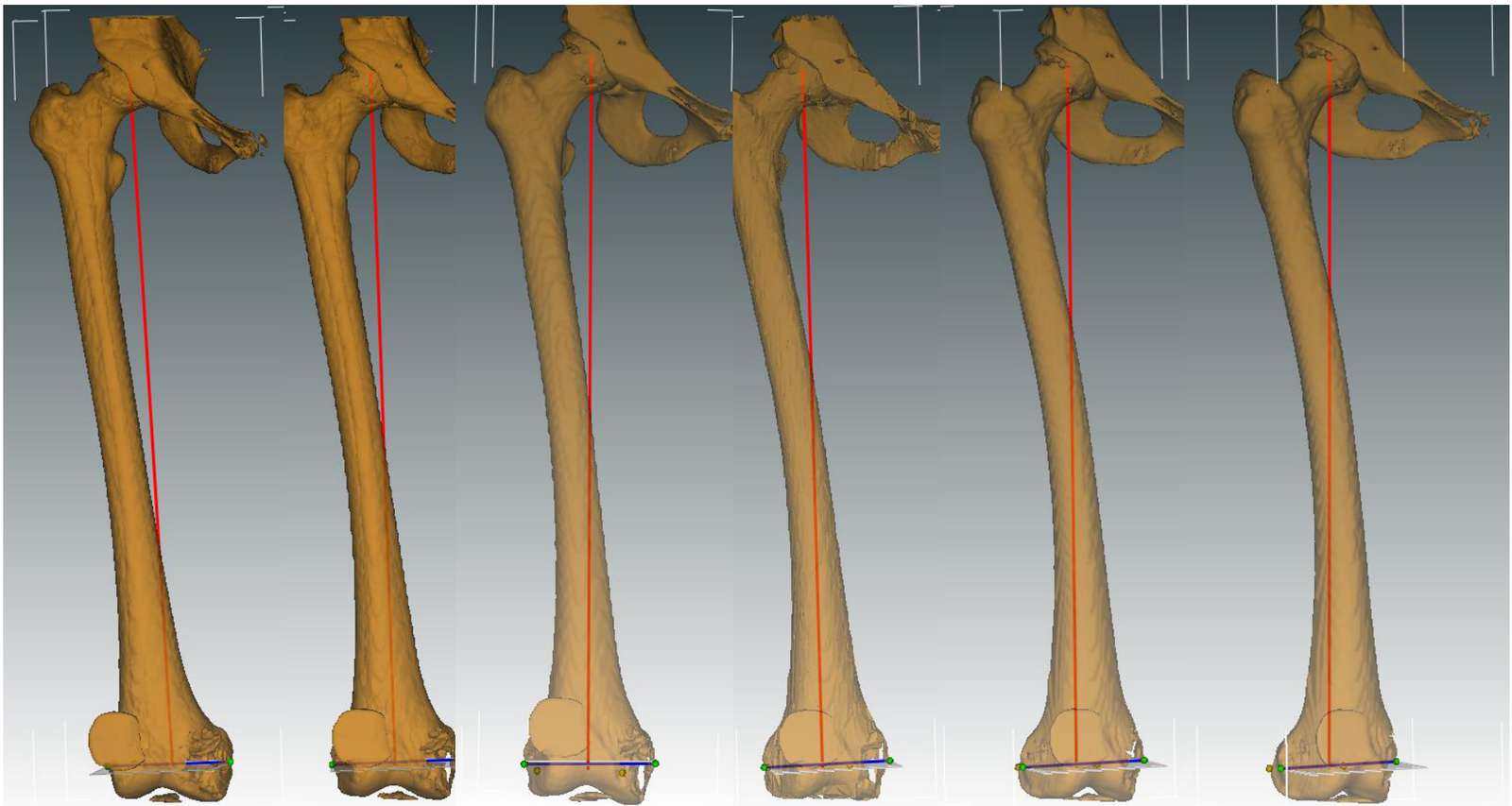
Three-dimensional planning allows the femoral and tibial morphology to be examined spatially, together with rotation, sagittal shape, and implant positioning. Even when CT segmentation and landmark detection are automated, the final plan should specify size, resection depth, alignment targets, and their rationale. Lan and colleagues’ retrospective comparison of 60 patients demonstrated the potential of AI-assisted 3D planning for size and axial alignment prediction. A small comparative study, however, cannot establish improved function or long-term implant survival for every patient.
AI that generates a plan, patient-specific instrumentation (PSI) that guides resection, and a robot that assists execution perform different roles. Connecting image-based bone planning with intraoperative soft-tissue balance requires checking the actual resection planes, flexion and extension gaps, and ligament condition, with plan adjustments when needed. CT time, cost, and radiation exposure, as well as registration errors, must also be considered. Alignment reproducibility, patient-reported pain and function, and long-term outcomes should each be evaluated.
Personalizing Care through Risk and Recovery Prediction
Personalized planning extends beyond implant selection. Research uses age, comorbidities, preoperative function, and patient-reported outcomes to predict discharge destination, length of stay, complications, and recovery trajectories. Booth and colleagues studied discharge destination after knee and hip arthroplasty using US NSQIP data, while Lopez and colleagues’ systematic review showed that model performance varies by prediction target. Discharge prediction and prediction of infection, pain, or function should not be interpreted through a single combined performance measure.
Predictions can inform patient counseling, rehabilitation resource allocation, and follow-up planning. Actual decisions also depend on caregiving support, the home environment, and patient preferences, which may be incompletely represented in the data. A high predicted risk does not establish the effectiveness of a particular intervention, so treatment and discharge timing should not be determined by predictions alone. Calibration against observed event rates in the local population and the effect of model use on recovery and care quality should be assessed.
Validation and Accountable Use in Clinical Workflows
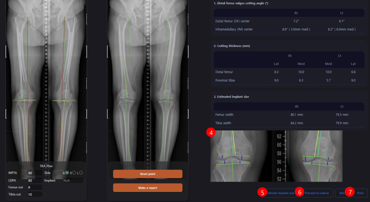
Clinical use requires checking measurements on the original images and surgeon review of a plan that links resection depth with implant size and position. Axes, landmarks, and segmentation errors must be correctable, and final approval and subsequent changes recorded. Connecting intraoperative anatomical and gap assessments with postoperative imaging and patient-reported outcomes allows discrepancies between the plan and actual results to be traced.
Meaningful validation should include different institutions, implant systems, and deformity patterns, while recording unprocessable images and cases requiring correction. Sizing evaluation should distinguish exact agreement from agreement within one size; alignment evaluation should assess differences from reference measurements and reproducibility. Risk models must use only information available at the prediction time. Ultimately, clinical value requires prospective assessment of total workload, including review and correction time, plan modification rates, complications, and patient-reported outcomes.
Related papers
- Park et al. A deep learning based automatic two-dimensional digital templating model for total knee arthroplasty (2024)
- Heller et al. Comparison of an AI-driven planning tool and manual radiographic measurements in total knee arthroplasty (2025)
- Lan et al. Reliable prediction of implant size and axial alignment in AI-based 3D preoperative planning for total knee arthroplasty (2024)
- Booth et al. Machine Learning to Predict Discharge Destination After Total Knee Arthroplasty and Total Hip Arthroplasty (2023)
- Lopez et al. Artificial Learning and Machine Learning Decision Guidance Applications in Total Hip and Knee Arthroplasty: A Systematic Review (2021)
- License for the cropped figure: Creative Commons Attribution 4.0