APKASS
Redefining Knee Osteoarthritis Through Imaging Biomarkers and AI
This lecture examines objective measurement, quantitative tracking of structural change, and individual prognosis in knee osteoarthritis. It connects joint space width, lower limb alignment, and MRI findings, and considers the clinical meaning of AI through validation in independent cohorts.
Select an image to view it at a larger size.
Understanding Osteoarthritis Through Quantitative Phenotypes
The Kellgren-Lawrence (K-L) grade provides a common language for describing the radiographic state of knee osteoarthritis. Reader disagreement near grade boundaries and limited sensitivity to small changes require separate consideration. AI-based grading addresses the consistency of interpretation, whereas continuous measurements of joint space width and alignment track structural changes within the same grade. Accumulating objective measurements allows both the current state and its change over time to be described.
The central research questions are how reproducibly osteoarthritis can be measured, which structures change first, and how those changes relate to the subsequent course. A 2024 study of automated K-L classification developed its model with Osteoarthritis Initiative (OAI) data and evaluated it in the independent Multicenter Osteoarthritis Study (MOST) dataset. This validation provides a basis for interpreting measurements and classifications across different patient populations. The relationship between symptoms, function, and structural imaging findings also requires evaluation when defining an individual patient’s phenotype.
Joint Space Biomarkers and Tissue-Specific Progression
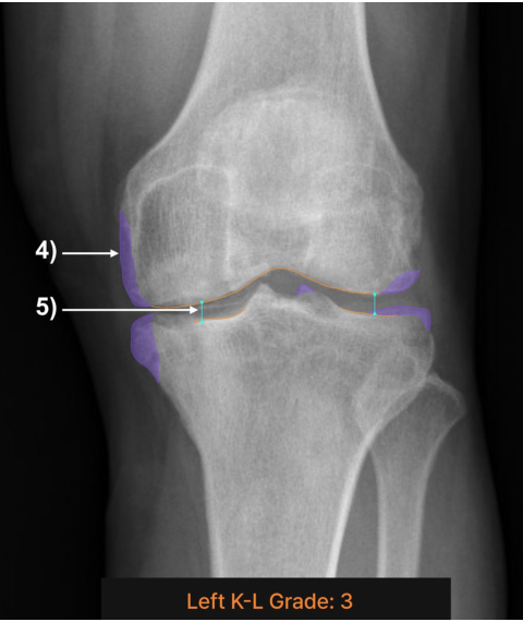
Orthogonal minimum joint space width (oJSW) is the narrowest joint space measured perpendicular to the tibial joint line within a standardized medial region. Evaluating it alongside fixed-location joint space widths explicitly defines the measurement location and its geometric relationship to the joint surface. The relevant study analyzed 15,313 knee radiographs collected in the OAI from baseline through 72 months, separately assessing discrimination of cross-sectional joint space narrowing and responsiveness to longitudinal change. Validating grading and change measurements separately clarifies the role of an imaging biomarker.
Joint space width should be interpreted in relation to cartilage and meniscal changes. A study of 3,710 knees with medial compartment osteoarthritis in seven years of MOST follow-up data found that cartilage damage preceded meniscal and bone changes centrally, whereas meniscal changes preceded cartilage and bone lesions posteriorly. Central femoral cartilage damage was observed even in K-L grade 0 and could appear before radiographic changes. These findings show that progression differs by compartment and tissue, supporting consideration of quantitative X-ray measures together with tissue information from MRI.
Reading the Rate of Deformity Through Serial Alignment Measurements
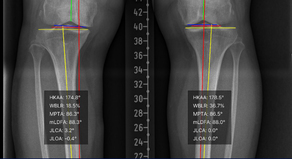
Lower limb alignment assessment measures the hip-knee-ankle angle (HKAA) together with the medial proximal tibial angle (MPTA), mechanical lateral distal femoral angle (mLDFA), and joint line convergence angle (JLCA). This combination describes the overall mechanical axis, the orientation of the femoral and tibial joint surfaces, and intra-articular convergence. A deep learning model recognizing anatomical landmarks was developed to calculate these angles automatically on full-leg radiographs. Agreement and test-retest reproducibility were evaluated against specialist reference measurements. Applying the same measures to follow-up images connects alignment status with the rate of change.
A longitudinal alignment study classified baseline HKAA as varus at 177° or less, neutral above 177° and below 183°, and valgus at 183° or more. A change of at least 0.50° per year defined accelerated progression. Baseline alignment, JLCA, and K-L grade were associated with subsequent accelerated alignment change. Recording a rate of change that accounts for the follow-up interval, alongside a single-time-point angle, is therefore central to this research. These associations represent prognostic information from an observational study; the effect of treatment on progression requires evaluation in studies of that treatment.
Models for Individual Prognosis and External Validation
A study predicting radiographic osteoarthritis progression evaluated K-L grade progression over 4-5 years in knees initially graded 0, 1, or 2. The model was developed in 5,966 OAI knees and validated in 3,392 MOST knees. Its selected features were baseline K-L grade, contralateral osteoarthritis, lateral and medial joint space narrowing, BMI, and total WOMAC score. External validation yielded a reported AUC of 0.76 and F1 score of 0.56. Performance measures are interpretable when accompanied by the predicted outcome, time horizon, and validation population. This study addressed radiographic grade progression; changes in pain or function and the need for surgery each require their own outcome definitions and validation.
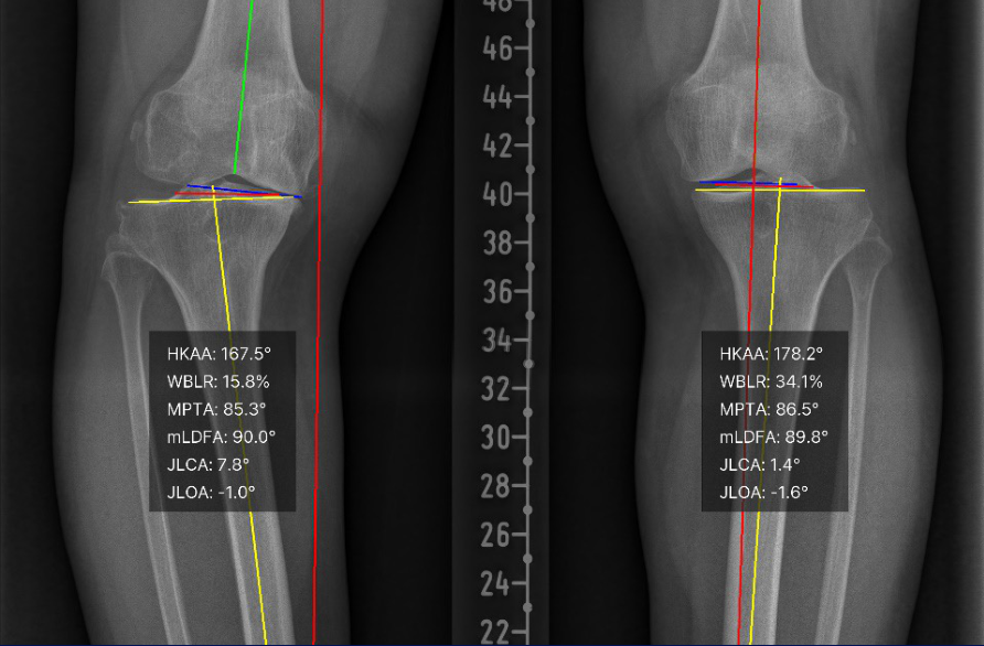
New-onset osteoarthritis in the contralateral knee is a separate prediction problem. A study of patients with unilateral osteoarthritis developed a model in 900 OAI participants and evaluated it in 453 MOST participants, predicting contralateral K-L grade 2 or higher after 4-5 years. The test dataset AUC was 0.69, and inputs included contralateral joint space narrowing and baseline grade. In the illustrated follow-up case, right-sided HKAA changed from 174.8° to 167.5° and JLCA from 3.2° to 7.8°. The case demonstrates what serial measurements can track, while the performance of an individual prognosis model is judged through evaluation in an independent cohort.
Connecting Objective Measurement to Clinical Decisions

Imaging analysis is extending from lower limb alignment and osteoarthritis grading to lateral knee measures, stress radiographs, MRI, and other tasks. The illustrated research-use interface organizes these tasks into separate modules. Specifying the anatomical target and output measures clarifies the purpose of repeated analysis. Evaluation for clinical use must likewise examine measurement reproducibility, applicability to images from other institutions, and prognostic validation for each particular task.
The purpose of quantification is to describe a patient’s current state, document the trajectory of change, and support judgment about the subsequent course. Connecting joint space and alignment measurements with symptoms, function, and the contralateral knee provides a more detailed understanding of the patient. Objective measurement, longitudinal follow-up, and validated individual prognosis are the research foundations of this approach. Its value in clinical care and public health should also be assessed through patient outcomes and costs, informing care adapted to different patterns of osteoarthritis progression.
Related papers
- Automated K-L Classification and Evaluation in an Independent Dataset (Lee et al., 2024)
- Orthogonal Minimum Joint Space Width and Assessment of Osteoarthritis Progression (Lee et al., 2025)
- Longitudinal X-ray–MRI Associations and Early Structural Changes (Lee et al., 2026)
- Automated Landmark Recognition and Angle Measurement on Full-Leg Radiographs (Jo et al., 2023)
- Joint Line Convergence Angle and Accelerated Alignment Progression (Kang et al., 2026)
- An Externally Validated Model for Radiographic Progression (Lee et al., 2025)
- Prediction of Contralateral Knee Osteoarthritis Development (Kim et al., 2025)