
2024 · Knee Surgery, Sports Traumatology, Arthroscopy
TEA 기준 3D CT 지표로 trochlear dysplasia를 정량화할 수 있을까?
활차 이형성군의 0°·15°·30° 평면에서 TG-TEA와 MP-MEC가 더 컸습니다(모두 P < 0.05).
원문 Figure 2
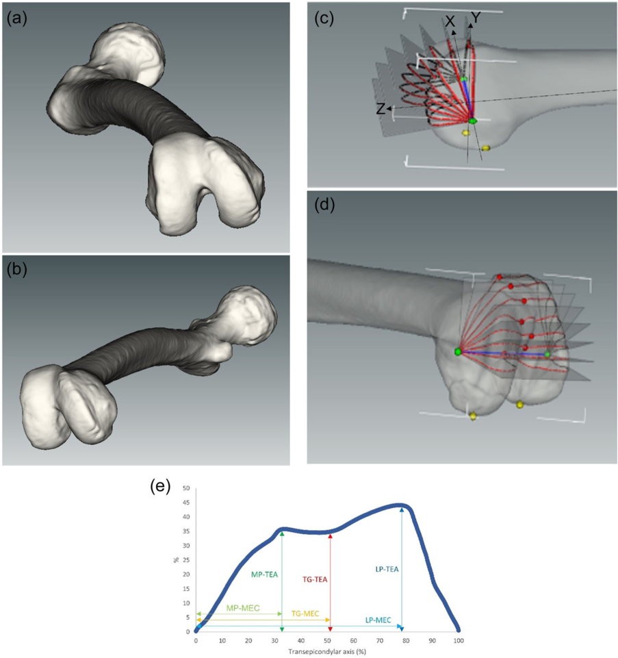 그림 크게 보기
3D CT로 활차의 모양을 재구성하고 일정한 축을 잡아 여러 높이의 단면을 비교합니다. 한 장의 측면 사진에서 보이는 분류를 넘어 어디가 얕고 좁은지 수치로 설명하려는 방법입니다.
Joon Young Lee, Sung Eun Kim, Oh Hyo Kwon, Youngjun Kim, Tae-geun Son, Hyuk-Soo Han, Du Hyun Ro. Shallow trochlear groove and narrow medial trochlear width at the proximal trochlea in patients with trochlear dysplasia: A three-dimensional computed tomography analysis. Knee Surgery, Sports Traumatology, Arthroscopy. 2024;32(6):1434–1445. https://doi.org/10.1002/ksa.12166.
원문 캡션
Figure 2
원문 Figure 3
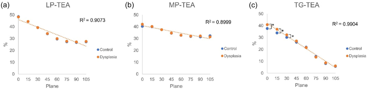 그림 크게 보기
단면의 각도가 달라질 때 활차 형상 지표가 어떻게 달라지는지 비교한 원문 도표입니다. 측정 위치가 달라지면 같은 이름의 지표도 다른 값을 낼 수 있습니다.
Joon Young Lee, Sung Eun Kim, Oh Hyo Kwon, Youngjun Kim, Tae-geun Son, Hyuk-Soo Han, Du Hyun Ro. Shallow trochlear groove and narrow medial trochlear width at the proximal trochlea in patients with trochlear dysplasia: A three-dimensional computed tomography analysis. Knee Surgery, Sports Traumatology, Arthroscopy. 2024;32(6):1434–1445. https://doi.org/10.1002/ksa.12166.
원문 캡션
Figure 3
대상과 방법
주요 결과
진료·수술에서 읽을 지점
해석의 제한
짧게 정리하면
이 연구가 확인한 것은 무엇인가요?
활차 이형성군의 0°·15°·30° 평면에서 TG-TEA와 MP-MEC가 더 컸습니다(모두 P < 0.05).
개별 환자의 치료 결정에 바로 적용할 수 있나요?
형태 연구입니다. Trochleoplasty·MPFL·TTO 기법의 효과를 비교하지 않았습니다. Dysplasia 20명으로 subtype 일반화에 제한이 있습니다.
Keywords · 주제어
- quantitative parameter
- three‐dimensional reconstruction
- trochlear dysplasia
- trochlear groove
- trochlear morphology
- Imaging, Three-Dimensional
- Tomography, X-Ray Computed
- Young Adult
Abstract · 원문 초록
Joon Young Lee, Sung Eun Kim, Oh Hyo Kwon, Youngjun Kim, Tae-Geun Son, Hyuk-Soo Han, Du Hyun Ro. Shallow trochlear groove and narrow medial trochlear width at the proximal trochlea in patients with trochlear dysplasia: A three-dimensional computed tomography analysis. Knee surgery, sports traumatology, arthroscopy : official journal of the ESSKA. 2024;32(6):1434-1445. doi:10.1002/ksa.12166.
PURPOSE
Although the Dejour classification is the primary classification system for evaluating trochlear dysplasia, concerns have been raised about its reliability owing to its qualitative criteria and challenges associated with obtaining accurate radiographs. This study aimed to quantify trochlear dysplasia using three-dimensional (3D) computed tomography (CT) reconstruction with novel parameters related to the transepicondylar axis (TEA).
METHODS
Sixty patients were enrolled, including 20 with trochlear dysplasia and 40 healthy controls. The 3D CT model was generated using the Materialise Interactive Medical Image Control System software. The following six parameters were measured in eight consecutive planes at 15° intervals (planes 0-105): the distance from the TEA to the most cortical point of the lateral condyle ('LP-TEA', where LP stands for lateral peak), medial condyle ('MP-TEA', MP for medial peak) and deepest point of the trochlea ('TG-TEA', TG for trochlear groove). The distances from the medial epicondyle (MEC) to the corresponding TEA points were measured ('LP-MEC', 'MP-MEC' and 'TG-MEC').
RESULTS
In the dysplasia group, TG-TEA (planes 0, 15 and 30) and MP-MEC (planes 0, 15 and 30) were significantly greater than those in the control group (all p < 0.05 for planes of TG-TEA and MP-MEC). For type A dysplasia, LP-MEC (plane 0) was greater than that in the control group. For type B dysplasia, the MP-MEC (planes 0 and 15) and TG-TEA (planes 0 and 15) were greater than those of the control group. For type D dysplasia, MP-MEC (planes 0, 15 and 30) and TG-TEA (planes 0 and 15) were elevated.
CONCLUSION
The 3D CT reconstruction analysis established a reproducible method for quantifying osseous trochlear morphology. Patients with trochlear dysplasia had a shallow TG and narrow medial trochlear width at tracking angles of 0°-30°. This finding corroborates the clinical manifestations of recurrent patellar instability that occur during early flexion.
LEVEL OF EVIDENCE
Level III.
원래 논문
Shallow trochlear groove and narrow medial trochlear width at the proximal trochlea in patients with trochlear dysplasia: A three-dimensional computed tomography analysis
Joon Young Lee, Sung Eun Kim, Oh Hyo Kwon, Youngjun Kim, Tae-Geun Son, Hyuk-Soo Han, Du Hyun Ro
Knee Surgery, Sports Traumatology, Arthroscopy · 2024