
2024 · Knee Surgery, Sports Traumatology, Arthroscopy
TEAを基準とした3D CT指標で、大腿骨滑車形成不全を定量化できるか?
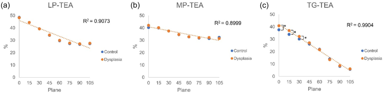
滑車形成不全群では、0°・15°・30°の断面でTG-TEAとMP-MECが大きくなっていました(すべてP < 0.05)。
原図Figure 2
 図を拡大する
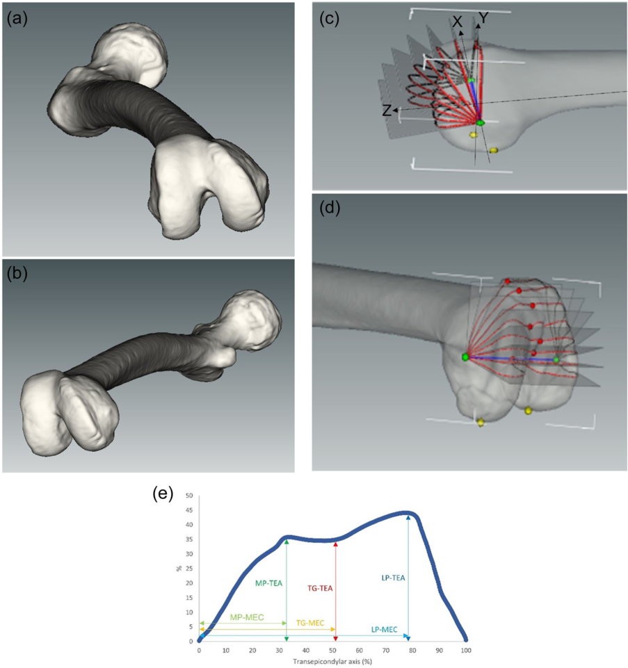
3DCTを使って弓の形を再現し安定した軸を採取して各高さのパーツを比較します 横写真で見られる分類よりも狭い場所を表す方法です
Joon Young Lee, Sung Eun Kim, Oh Hyo Kwon, Youngjun Kim, Tae-geun Son, Hyuk-Soo Han, Du Hyun Ro. Shallow trochlear groove and narrow medial trochlear width at the proximal trochlea in patients with trochlear dysplasia: A three-dimensional computed tomography analysis. Knee Surgery, Sports Traumatology, Arthroscopy. 2024;32(6):1434–1445. https://doi.org/10.1002/ksa.12166.
原著の図の説明
Figure 2
原図Figure 3
 図を拡大する
角が変化するときに gradientパターンがどのように変わるのかを比較します 測定位置が異なる場合,同じ名前を持つ指標は別の値を生成することができます.
Joon Young Lee, Sung Eun Kim, Oh Hyo Kwon, Youngjun Kim, Tae-geun Son, Hyuk-Soo Han, Du Hyun Ro. Shallow trochlear groove and narrow medial trochlear width at the proximal trochlea in patients with trochlear dysplasia: A three-dimensional computed tomography analysis. Knee Surgery, Sports Traumatology, Arthroscopy. 2024;32(6):1434–1445. https://doi.org/10.1002/ksa.12166.
原著の図の説明
Figure 3
対象と方法
主な結果
手術に関する読書のポイント
解釈の限界
簡単に説明しましょう
この研究で何が確認されましたか?
滑車形成不全群では、0°・15°・30°の断面でTG-TEAとMP-MECが大きくなっていました(すべてP < 0.05)。
治療決定に直接適用できるか?
形態を調べた研究です。Trochleoplasty・MPFL・TTOの手技の効果は比較していません。形成不全の患者さんは20人で、サブタイプへの一般化には限界があります。
Keywords・キーワード
- quantitative parameter
- three‐dimensional reconstruction
- trochlear dysplasia
- trochlear groove
- trochlear morphology
- Imaging, Three-Dimensional
- Tomography, X-Ray Computed
- Young Adult
Abstract・原文の抄録
Joon Young Lee, Sung Eun Kim, Oh Hyo Kwon, Youngjun Kim, Tae-Geun Son, Hyuk-Soo Han, Du Hyun Ro. Shallow trochlear groove and narrow medial trochlear width at the proximal trochlea in patients with trochlear dysplasia: A three-dimensional computed tomography analysis. Knee surgery, sports traumatology, arthroscopy : official journal of the ESSKA. 2024;32(6):1434-1445. doi:10.1002/ksa.12166.
PURPOSE
Although the Dejour classification is the primary classification system for evaluating trochlear dysplasia, concerns have been raised about its reliability owing to its qualitative criteria and challenges associated with obtaining accurate radiographs. This study aimed to quantify trochlear dysplasia using three-dimensional (3D) computed tomography (CT) reconstruction with novel parameters related to the transepicondylar axis (TEA).
METHODS
Sixty patients were enrolled, including 20 with trochlear dysplasia and 40 healthy controls. The 3D CT model was generated using the Materialise Interactive Medical Image Control System software. The following six parameters were measured in eight consecutive planes at 15° intervals (planes 0-105): the distance from the TEA to the most cortical point of the lateral condyle ('LP-TEA', where LP stands for lateral peak), medial condyle ('MP-TEA', MP for medial peak) and deepest point of the trochlea ('TG-TEA', TG for trochlear groove). The distances from the medial epicondyle (MEC) to the corresponding TEA points were measured ('LP-MEC', 'MP-MEC' and 'TG-MEC').
RESULTS
In the dysplasia group, TG-TEA (planes 0, 15 and 30) and MP-MEC (planes 0, 15 and 30) were significantly greater than those in the control group (all p < 0.05 for planes of TG-TEA and MP-MEC). For type A dysplasia, LP-MEC (plane 0) was greater than that in the control group. For type B dysplasia, the MP-MEC (planes 0 and 15) and TG-TEA (planes 0 and 15) were greater than those of the control group. For type D dysplasia, MP-MEC (planes 0, 15 and 30) and TG-TEA (planes 0 and 15) were elevated.
CONCLUSION
The 3D CT reconstruction analysis established a reproducible method for quantifying osseous trochlear morphology. Patients with trochlear dysplasia had a shallow TG and narrow medial trochlear width at tracking angles of 0°-30°. This finding corroborates the clinical manifestations of recurrent patellar instability that occur during early flexion.
LEVEL OF EVIDENCE
Level III.
オリジナル紙だ
Shallow trochlear groove and narrow medial trochlear width at the proximal trochlea in patients with trochlear dysplasia: A three-dimensional computed tomography analysis
Joon Young Lee, Sung Eun Kim, Oh Hyo Kwon, Youngjun Kim, Tae-Geun Son, Hyuk-Soo Han, Du Hyun Ro
Knee Surgery, Sports Traumatology, Arthroscopy · 2024