2022 · Journal of Clinical Medicine
하지 전장 X-ray의 근육 영역을 이용해 수술 전 근감소증을 선별할 수 있을까요?
분할 모델의 시험군 IoU는 0.926이었습니다. PMV와 골격근지수의 상관계수는 0.654였습니다.
원문 Figure 1 · X-ray의 근육 영역을 분리합니다
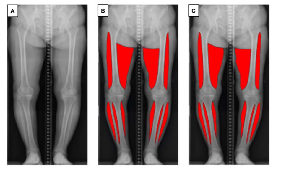 그림 크게 보기
A는 원본 영상, B는 연구자가 표시한 근육 영역, C는 모델이 예측한 영역입니다. X-ray에서 근육 형태를 정량화하는 방법을 보여줍니다.
Doohyun Hwang, Sungho Ahn, Yong-Beom Park, Seong Hwan Kim, Hyuk-Soo Han, Myung Chul Lee, Du Hyun Ro. Journal of Clinical Medicine (2022). DOI: 10.3390/jcm11133612
연구에서 묻는 질문
대상과 방법
주요 결과
진료와 수술에서 읽을 지점
해석의 제한
짧게 정리하면
어떤 진료 질문과 연결되나요?
수술 전에 이미 촬영한 영상과 혈액검사에서 근감소증 위험을 살펴볼 수 있다는 연구입니다. 영양 상태와 근력·보행 평가를 함께 확인할 때 활용 가능성을 검토할 수 있습니다.
Keywords · 주제어
- deep learning
- osteoarthritis
- plain radiograph
- sarcopenia
- screening
- segmentation
- total knee arthroplasty
- AI
Abstract · 원문 초록
Doohyun Hwang, Sungho Ahn, Yong-Beom Park, Seong Hwan Kim, Hyuk-Soo Han, Myung Chul Lee, Du Hyun Ro. Deep Learning-Based Muscle Segmentation and Quantification of Full-Leg Plain Radiograph for Sarcopenia Screening in Patients Undergoing Total Knee Arthroplasty. Journal of clinical medicine. 2022;11(13):3612. doi:10.3390/jcm11133612.
Sarcopenia, an age-related loss of skeletal muscle mass and function, is correlated with adverse outcomes after some surgeries. Here, we present a deep-learning-based model for automatic muscle segmentation and quantification of full-leg plain radiographs. We illustrated the potential of the model to predict sarcopenia in patients undergoing total knee arthroplasty (TKA). A U-Net-based deep learning model for automatic muscle segmentation was developed, trained and validated on the plain radiographs of 227 healthy volunteers. The radiographs of 403 patients scheduled for primary TKA were reviewed to test the developed model and explore its potential to predict sarcopenia. The proposed deep learning model achieved mean IoU values of 0.959 (95% CI 0.959-0.960) and 0.926 (95% CI 0.920-0.931) in the training set and test set, respectively. The fivefold AUC value of the sarcopenia classification model was 0.988 (95% CI 0.986-0.989). Of seven key predictors included in the model, the predicted muscle volume (PMV) was the most important of these features in the decision process. In the preoperative clinical setting, wherein laboratory tests and radiographic imaging are available, the proposed deep-learning-based model can be used to screen for sarcopenia in patients with knee osteoarthritis undergoing TKA with high sarcopenia screening performance.
© 2022 by the authors.
원래 논문
Deep Learning-Based Muscle Segmentation and Quantification of Full-Leg Plain Radiograph for Sarcopenia Screening in Patients Undergoing Total Knee Arthroplasty
Doohyun Hwang, Sungho Ahn, Yong-Beom Park, Seong Hwan Kim, Hyuk-Soo Han, Myung Chul Lee, Du Hyun Ro
Journal of Clinical Medicine · 2022